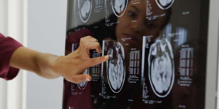
![[Adobe Stock]](https://sp-ao.shortpixel.ai/client/to_webp,q_glossy,ret_img,w_750,h_375/https://www.psypost.org/wp-content/uploads/2025/03/brain-MRI-scans-750x375.jpg)
A new study published in Brain Imaging and Behavior has uncovered differences in brain function and neurotransmitter systems in men with lifelong premature ejaculation. Using advanced brain imaging, the researchers found that individuals with this condition showed altered activity in several brain regions related to emotion, sensation, and visual processing. These brain changes were also linked to key neurotransmitter systems that regulate reward, arousal, and muscle control.
Lifelong premature ejaculation is a common male sexual disorder marked by consistent early ejaculation, often within one minute of sexual activity. It can cause significant distress, affect self-esteem, and create tension in relationships. Despite its prevalence—affecting around 3% of men in China and up to 5% globally—the underlying brain mechanisms are still not fully understood. Diagnoses typically rely on self-reported measures, and objective biological markers are lacking.
To address this gap, researchers in China set out to investigate how the brains of men with lifelong premature ejaculation differ from those of men without the condition. They aimed to find measurable patterns of brain activity that could help explain why some men are affected. Their approach focused on resting-state brain function using a method called percent amplitude of fluctuation, or PerAF. This technique measures how much blood flow-related activity varies in different regions of the brain when a person is not engaged in any specific task.
The study involved 81 men—46 with lifelong premature ejaculation and 35 healthy controls. All participants were screened for mental and physical health and were not taking medication. Brain scans were conducted using a 3.0 Tesla MRI scanner while participants rested with their eyes closed. The researchers then analyzed the brain imaging data to identify regions where brain activity differed between the two groups. They also compared the imaging findings with clinical measures of sexual function, anxiety, and depression.
The researchers found that men with premature ejaculation had increased brain activity in several key regions. These included the middle cingulate cortex, supramarginal gyrus, Rolandic operculum, hippocampus and parahippocampal area, and the insula. These areas are known to be involved in emotional regulation, sensory integration, and bodily awareness. At the same time, reduced activity was observed in the precuneus, inferior temporal cortex, and parts of the occipital lobe—regions linked to self-referential thinking, visual imagery, and attention.
To test whether these brain differences could help distinguish between individuals with and without premature ejaculation, the researchers conducted a receiver operating characteristic analysis. This statistical method estimates how accurately a variable—such as brain activity in a specific region—can classify a condition. The analysis showed that some of the altered brain regions, especially the middle cingulate cortex and hippocampus, were moderately effective in distinguishing the affected group from controls.
The study also found meaningful connections between brain activity and clinical symptoms. Higher activity in the insula was associated with greater depression scores, while lower activity in the precuneus was linked to poorer sexual function. These findings suggest that emotional and cognitive processes may play a role in how premature ejaculation develops and persists.
To explore the biological underpinnings of these brain changes, the researchers investigated whether the altered activity was related to known patterns of neurotransmitter distribution. They used a brain mapping tool called JuSpace, which compares imaging data with maps of neurotransmitter systems, including dopamine, serotonin, norepinephrine, and acetylcholine. The analysis showed that brain activity patterns in men with premature ejaculation were significantly correlated with areas rich in dopamine D2 receptors, serotonin transporters, norepinephrine transporters, and vesicular acetylcholine transporters.
These neurotransmitters are involved in a wide range of processes relevant to sexual function. Dopamine plays a key role in motivation, reward, and motor control. Serotonin is involved in mood regulation and is known to delay ejaculation when increased. Norepinephrine affects arousal and muscle contractions, while acetylcholine is important for communication between nerves and muscles. Imbalances in these systems may help explain why some individuals experience rapid ejaculation and emotional distress.
The researchers suggest that premature ejaculation may be partly driven by a failure to balance sexual arousal and inhibitory control. This could result from dysregulation in the brain’s reward and emotional circuits, leading to faster ejaculation that is difficult to consciously control. Some scientists have proposed that premature ejaculation may resemble impulse control disorders, which share similar disruptions in neurotransmitter systems. Other studies have linked premature ejaculation to traits like anxiety, impaired self-regulation, and heightened sensitivity to sexual stimuli.
Despite these insights, the study has some limitations. It was based on a relatively small sample and used a cross-sectional design, which means it cannot determine whether the brain differences cause the condition or are a result of it. The researchers also relied solely on functional imaging data, without combining it with structural scans or other biological measures. Future studies with larger and more diverse samples, as well as multimodal imaging approaches, may offer a more complete picture.
The study, “Abnormal percent amplitude of fluctuation in patients with lifelong premature ejaculation is associated with neurotransmitter profiles,” was authored by Jiarui Yuan, Pinxiao Wang, Dingxin Nie, Wanxiang Zheng, Kepu Liu, Jianyong Feng, Yuntao Zhang, Yanzhu Wang, Junjun Gao, and Ming Gao.


