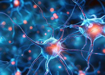
![This image shows the prefrontal cortex of a mouse injected with scFLARE2 (purple) which drives the expression of a light-responsive channel in psychedelic-activated neurons (green or red). This allows researchers to visualize and artificially reactive these neurons at a later time point. [Christina Kim, UC Davis]](https://sp-ao.shortpixel.ai/client/to_webp,q_glossy,ret_img,w_750,h_375/https://www.psypost.org/wp-content/uploads/2025/05/neurons-2-750x375.jpg)
A new study published in Science suggests that the anxiety-reducing effects of psychedelics can be reproduced by stimulating a specific group of brain cells, without triggering hallucination-like behaviors. In experiments with mice, researchers identified a set of neurons in the medial prefrontal cortex that are activated by a psychedelic compound and linked to decreased anxiety. When these neurons were reactivated using light the next day, the mice still showed less anxiety — even though the drug was no longer present in their system and the head-twitch responses associated with psychedelic-like experiences had faded.
Psychedelic compounds have shown therapeutic promise for mood and anxiety disorders, especially in people who do not respond to traditional treatments. However, the hallucinogenic effects of these substances raise safety concerns and limit their widespread clinical use. While some newer compounds aim to remove the hallucinogenic effects chemically, researchers in this study pursued a different strategy: pinpointing the brain circuits responsible for the therapeutic benefit and testing whether stimulating those circuits alone could mimic the desired effects.
To explore this idea, the research team at the University of California, Davis administered the psychedelic compound DOI (2,5-dimethoxy-4-iodoamphetamine) to mice. DOI acts on serotonin receptors in the brain and has been previously shown to reduce anxiety-like behaviors in animals. The team first tracked the presence of the drug in the brain using a fluorescent biosensor that lights up when serotonin receptors are activated. They found that the drug peaked in the brain around 30 minutes after injection and was largely undetectable after four hours.
To measure behavior, the researchers used two standard tests: the marble burying test and the elevated plus maze. In the marble burying test, anxious mice tend to compulsively bury marbles in their bedding. In the elevated plus maze, anxious mice avoid exploring the open arms of a raised cross-shaped maze. Mice given DOI buried fewer marbles and spent more time exploring the open arms, indicating reduced anxiety. Importantly, while the hallucinogenic-like head twitch response appeared only within the first few hours after dosing, the reduction in anxiety-like behavior lasted beyond that window.
“It is well known that in mice, psychedelics induce reduced marble burying and promote exploration of the open arms of the plus maze,” said Christina Kim, the study’s corresponding author and an assistant professor of neurology, core member of the Center for Neuroscience. “But there is also an intoxicating or hallucinogenic-like effect, which can be measured through head twitches in mice.”
The team next examined how DOI affected neuronal activity in the medial prefrontal cortex, a region involved in mood and decision-making. Using a fluorescent calcium sensor and a miniature lens implanted in the brain, they monitored real-time activity in individual neurons. They found that about half of the neurons in this region increased their activity after DOI administration, while others were either unaffected or suppressed.
To identify the specific neurons involved in this process, the researchers used a molecular tagging tool called scFLARE2. This system allowed them to genetically label the cells that were active during a brief window following DOI administration. Later, they could reactivate just those tagged cells using optogenetics—a technique that uses light to control genetically modified cells.
When the researchers reactivated the DOI-tagged neurons 24 hours after the drug was given, the mice again showed reduced anxiety-like behavior in both the marble burying and elevated plus maze tests. At that point, DOI was no longer present in the brain, and the mice did not show head twitch behaviors. This indicates that the therapeutic effects could be recreated through specific circuit activation without triggering behaviors linked to hallucinogenic effects.
To better understand which types of neurons were involved, the team performed single-nucleus RNA sequencing on the tagged cells. They found that DOI activated a mixture of excitatory and inhibitory neurons, especially within certain layers of the prefrontal cortex. Some of the activated neurons expressed the serotonin receptor targeted by DOI, while others did not. This pattern suggests that DOI initially activates a specific subset of receptor-expressing cells, which then go on to recruit additional downstream neurons to produce behavioral effects.
Interestingly, when the researchers blocked serotonin receptors using an antagonist before DOI administration, they could prevent both the behavioral effects and the ability to later reactivate the anxiety-reducing response. This confirms that initial receptor activation is necessary to engage the network of cells responsible for the therapeutic effects.
The study also explored whether optogenetic reactivation of these cells produced any side effects similar to those seen during psychedelic exposure. The mice showed no increase in head twitch behaviors during reactivation, reinforcing the idea that the anxiety-reducing effects of the drug and its hallucinogenic effects can be separated at the circuit level.
“In the past, we did this using chemistry by making new compounds, but here we focused on identifying the circuits responsible for the effects, and it does seem that they are distinct,” said study co-author David E. Olson, director of the Institute for Psychedelics and Neurotherapeutics (IPN) and a professor of chemistry and of biochemistry and molecular medicine. “This is an important mechanistic study that validates our earlier results.”
While the findings offer new insights into how psychedelics work in the brain, the researchers note that their study has some limitations. The work was conducted in mice, and while the marble burying and maze tests are widely used to study anxiety, they cannot fully capture the human experience. Additionally, the tagging system used in the study only labels activated neurons and not those that are suppressed, which may also play a role in shaping behavior.
Another limitation is the use of DOI itself. While it shares many properties with other classical psychedelics, it is not being considered for human therapeutic use. “While DOI is a potent psychedelic, it is not being explored as a potential therapeutic drug in the clinic. Thus the findings here are focused on dissecting the basic circuit mechanisms of this important class of drugs,” Kim said.
The researchers suggest that different behavioral effects may arise from distinct populations of neurons, even if they are activated by the same compound. Future studies will aim to further map these circuits and determine how changes in activity, plasticity, and gene expression contribute to lasting improvements in mood and anxiety.
“Understanding which neural circuits psychedelics activate to elicit their effects is the kind of basic science needed to ultimately develop targeted therapeutics with better safety profiles,” Olson said.
The study, “Isolation of psychedelic-responsive neurons underlying anxiolytic behavioral states,” was authored by J. Muir, S. Lin, I. K. Aarrestad, H. R. Daniels, J. Ma, L. Tian, D. E. Olson, and C. K. Kim.


