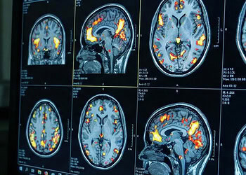
![[Adobe Stock]](https://sp-ao.shortpixel.ai/client/to_webp,q_glossy,ret_img,w_750,h_375/https://www.psypost.org/wp-content/uploads/2024/10/brain-scans-750x375.jpg)
A new study published in Scientific Reports provides a detailed model of how the human brain develops during the transition from the womb to early infancy. The findings indicate that distinct growth patterns for different brain tissues and sex-based differences in brain volume are established between mid-pregnancy and the first weeks of life. This research offers a continuous view of how the brain expands during a foundational period that was previously difficult to map.
The perinatal period involves rapid biological changes that establish the core architecture of the human brain. This phase includes the processes where cells proliferate, migrate to their correct locations, and begin forming complex connections. Scientists have often studied prenatal development and postnatal development separately because of the technical challenges involved in imaging fetuses compared to newborns. This separation has historically made it difficult to understand exactly how growth trajectories evolve as a fetus becomes an infant.
To bridge this gap, a research team led by the University of Cambridge aimed to create a unified model of early brain growth. Yumnah T. Khan, a PhD student at the Autism Research Centre at the University of Cambridge, led the investigation. The team sought to determine when specific tissues dominate growth and when sex differences in brain size first appear. By combining data from before and after birth, they hoped to capture the dynamic nature of brain structural changes.
The researchers utilized data from the Developing Human Connectome Project, which is a large-scale initiative designed to map brain connectivity. The final dataset included 798 magnetic resonance imaging scans collected from 699 unique individuals. These participants included 263 fetuses scanned while in the womb and 535 newborns.
The sample consisted of 380 males and 319 females. The scans covered a developmental window ranging from just over 21 weeks to nearly 45 weeks after conception. This allowed the team to track changes across the second and third trimesters of pregnancy and into the first month after birth.
The team used advanced statistical modeling to chart the volume of different brain tissues against the age of the individuals. They applied corrections to account for the natural variance that increases as infants grow older. The analysis focused on total brain volume as well as specific compartments like gray matter, white matter, and cerebrospinal fluid.
The analysis revealed that the total volume of the brain grows at an increasing rate leading up to birth. When the researchers accounted for the exact age at the time of the scan, they observed a slight slowing of this growth rate in the weeks immediately following birth. This suggests the most rapid expansion occurs just before and shortly after delivery.
Different types of brain tissue followed their own unique timelines. White matter, which forms the connections between brain cells, was the primary driver of growth during mid-pregnancy. However, its proportional contribution to the total brain size decreased over time. This suggests the brain prioritizes establishing core connectivity pathways early in gestation.
In contrast, gray matter, which contains the cell bodies of neurons and is involved in processing information, became the dominant driver of growth during late pregnancy and the postnatal period. This shift indicates a transition from laying down connections to the proliferation and maturation of processing centers. The rapid growth of gray matter likely supports the development of sensory and motor abilities needed for survival after birth.
The study also looked at deep brain structures known as subcortical regions. These areas, such as the amygdala and thalamus, showed an earlier peak in their growth rates compared to the outer layer of the brain, the cortex. The cortex is typically associated with higher-level cognitive functions.
The finding that subcortical structures mature faster aligns with the understanding that regions responsible for basic physiological and sensory functions develop before those involved in complex thought. The researchers observed that the cerebellum, a region critical for motor control, showed exponential growth throughout the studied period. This rapid expansion likely facilitates the early coordination required for an infant’s movements.
A major component of the analysis involved comparing brain development between males and females. The data showed that, on average, males experienced greater increases in brain volume as they aged compared to females. This difference was observable across the entire brain and within specific regions.
The researchers found that these sex differences were generally linear, meaning males consistently showed faster growth. This provides evidence that sex differences in brain structure are not solely a result of social or environmental influences after birth. Instead, biological factors present during pregnancy appear to initiate these divergence patterns.
While males exhibited faster overall growth, the shape of the growth trajectories was largely similar between the sexes. Both males and females followed the same general patterns of tissue expansion. However, there were specific exceptions in regional development.
For example, parts of the temporal lobe showed more pronounced gray matter increases in males. Additionally, the team identified a distinct growth pattern in the left anterior cingulate gyrus. In this region, males showed an S-shaped growth curve, whereas females showed a linear trajectory.
The study faces certain limitations regarding the available data. The scans for fetuses did not begin until after 21 weeks of gestation, leaving the first half of pregnancy unmapped in this analysis. Additionally, the number of scans available for younger fetuses was smaller than for older infants, which could impact the precision of the early growth models.
The researchers also noted technical differences between how fetal and neonatal scans were acquired. Although the same scanner was used, the settings had to be adjusted for the different environments of the womb and the nursery. This could potentially introduce variations in the measurements, though the team observed strong continuity in the data.
While the study documents when sex differences emerge, it does not confirm the biological mechanisms causing them. The authors suggest that prenatal hormones like testosterone likely play a role. Male fetuses are exposed to a surge of testosterone between 14 and 18 weeks of gestation.
The timing of the observed structural differences, appearing after 18 weeks, corresponds with the aftermath of this hormonal surge. Future research will need to directly investigate the link between hormone levels and these structural changes to confirm causality. The researchers emphasize that understanding these typical growth trajectories provides a baseline for identifying atypical development.
This baseline could eventually help explain why certain neurodevelopmental conditions are more common in one sex than the other. For instance, autism is diagnosed more frequently in males. Understanding if and how early brain overgrowth relates to these conditions remains a priority for the field.
The team calls for further longitudinal studies to validate these findings over longer periods. Following the same individuals from pregnancy through childhood would provide even stronger evidence for these developmental patterns. The current study represents a significant step toward a complete map of early human brain development.
The study, “Mapping brain growth and sex differences across prenatal to postnatal development,” was authored by Yumnah T. Khan, Alex Tsompanidis, Marcin A. Radecki, Carrie Allison, Meng-Chuan Lai, Richard A. I. Bethlehem and Simon Baron-Cohen.


