![[Adobe Stock]](https://sp-ao.shortpixel.ai/client/to_webp,q_glossy,ret_img,w_750,h_375/https://www.psypost.org/wp-content/uploads/2025/06/neuron-750x375.jpg)
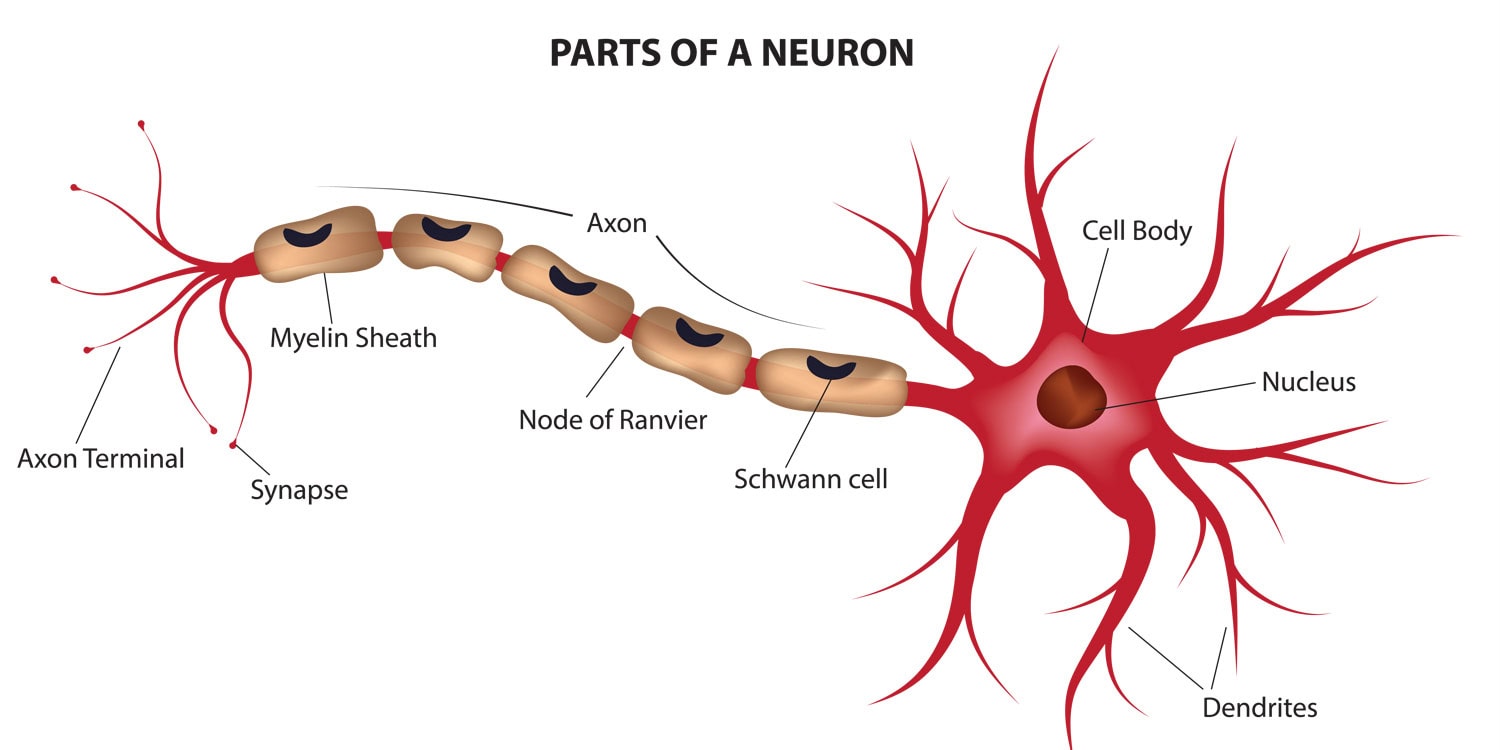
A new study published in Nature Metabolism suggests that intense endurance exercise, such as running a marathon, may cause a temporary reduction in myelin content in the brain. Using advanced magnetic resonance imaging (MRI) techniques, researchers observed that myelin—a fatty substance that insulates nerve fibers and supports brain function—was significantly reduced shortly after marathon running, but returned to normal levels within two months. These findings point to a previously unrecognized form of brain plasticity, where myelin may serve as an energy reserve under extreme metabolic conditions.
The motivation for the study came from a growing body of evidence showing that myelin, which makes up the white matter in the brain, is not only important for nerve conduction but may also play a role in brain metabolism. Myelin is composed mainly of lipids, and recent animal studies have suggested that in times of energy shortage, the body might tap into these lipid-rich structures to support brain function. The researchers wanted to test whether this could also happen in humans undergoing intense physical exertion, such as marathon runners who often experience significant depletion of their body’s primary energy sources during a race.
“I am a runner and have been studying myelin biology for three decades. In seeking a framework to explore the notion that brain myelin serves as an energy source, I considered the metabolic stress of marathon running as a potential way to test this hypothesis,” explained study author Carlos Matute, a professor at the University of the Basque Country and CIBERNED-Instituto Carlos III.
To investigate this possibility, the research team recruited 10 experienced marathon runners between the ages of 45 and 73. These volunteers, who were in good health and did not receive any financial compensation, participated in various marathon events including city and mountain races. Each runner underwent a series of MRI scans: once within 48 hours before their race, again 24 to 48 hours after, and for a subset of participants, at two weeks and two months after completing the marathon.
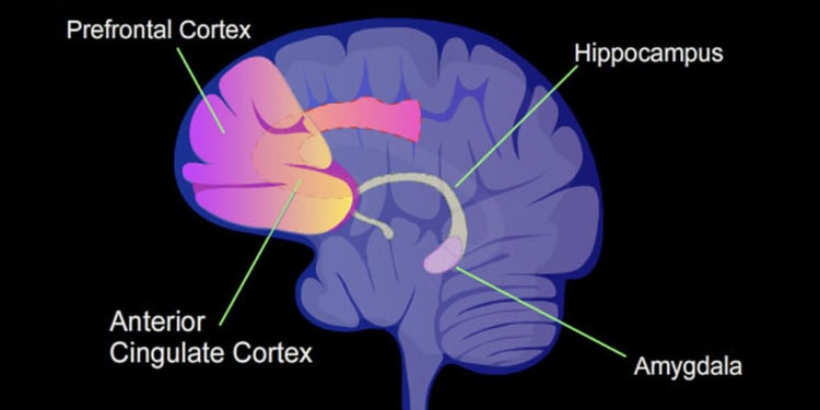
Initial scans taken before the race showed consistent MWF patterns across participants, with normal individual variability. After the race, however, the MRI data revealed a significant reduction in MWF in multiple white matter regions of the brain, particularly in areas linked to motor coordination, sensory processing, and emotional regulation. Some of the most affected regions included the corticospinal tract, pontine crossing tract, and cerebellar peduncles—tracts involved in movement and balance.
On average, the MWF dropped by as much as 28% in certain tracts within two days of completing the marathon. Notably, the reductions were observed in both hemispheres of the brain and did not occur uniformly across all regions. The changes were especially pronounced in highly myelinated white matter areas, while most grey matter regions remained unaffected, likely due to their lower baseline myelin content and limited sensitivity of the imaging method in those regions.
Importantly, the researchers ruled out several alternative explanations for the MWF changes. Dehydration, for instance, could alter water distribution in the brain and skew imaging results, but no significant changes in brain volume or regional water content were detected. Other potential confounding factors, such as brain swelling (edema), iron level fluctuations, and MRI signal orientation effects, were also considered unlikely contributors based on previous studies and the design of this research.
Two weeks after the race, follow-up scans showed that myelin levels were beginning to rebound but had not yet returned to pre-race levels. By the two-month mark, however, MWF values had fully recovered in all previously affected brain regions. This reversible pattern indicates that the reduction in MWF was not a sign of long-term damage but rather a temporary adaptation—what the authors describe as a form of “metabolic myelin plasticity.”
“The main limitation was technical—I initially believed it would be difficult to detect changes in myelin content,” Matute told PsyPost. “So it was surprising to observe that myelin levels can rapidly decrease within just 3 to 4 hours, the typical duration of a marathon, and then recover within a few weeks. This finding highlights a key point: the brain is more dynamic and plastic than previously thought. Another unexpected discovery was that these changes occur across a wide age range; the runners in our study were between 45 and 73 years old.”
The findings support the “idea that the brain harbors a substantial fat depot—myelin—which could potentially be used to fuel its activity,” Matute said. During a marathon, the body rapidly depletes its carbohydrate stores and turns to fat for energy. The brain may follow a similar strategy, mobilizing lipid reserves stored in the myelin sheath to sustain its function. Animal research has shown that glial cells—the support cells that produce myelin—can metabolize fatty acids through a process known as β-oxidation to generate energy during periods of glucose shortage. This metabolic flexibility may help protect nerve fibers and maintain communication within the brain under extreme physical stress.
Although the observed reduction in myelin was modest and transient, the implications are significant. If myelin lipids can be used as an emergency energy source, this may shed light on how the brain copes with metabolic challenges beyond exercise, including malnutrition or neurological diseases characterized by impaired energy balance. In fact, studies have shown that myelin integrity is affected in conditions like anorexia nervosa and neurodegenerative diseases, raising questions about whether similar metabolic mechanisms are involved.
There are some limitations to the study. The sample size was small, with only 10 participants, and most follow-up data were limited to subsets of this group. The researchers also could not directly measure myelin at the cellular level, as no non-invasive tools currently exist to do so with complete accuracy. MWF, while reliable, is considered a semiquantitative measure and can be influenced by factors unrelated to myelin degradation, such as minor changes in water distribution or tissue composition.
Future research will need to confirm these findings in larger and more diverse populations. The authors also plan to investigate whether the temporary loss of myelin observed after a marathon has any short-term effects on brain function, cognition, or mood. These follow-up studies could help determine whether changes in myelin content translate into measurable differences in how the brain performs after intense physical activity.
“The next steps involve investigating whether changes in brain function and cognition accompany the observed alterations in myelin,” Matute explained. “In the long term, our goal is to uncover the cellular and molecular mechanisms that mediate myelin consumption as an energy source for the brain—and its subsequent recovery. Gaining insight into these questions may aid in the development of therapies for patients with demyelinating diseases such as multiple sclerosis, as well as for addressing the impact of age-related myelin decline on brain function and cognition.”
The study, “Reversible reduction in brain myelin content upon marathon running,” was authored by Pedro Ramos-Cabrer, Alberto Cabrera-Zubizarreta, Daniel Padro, Mario Matute-González, Alfredo Rodríguez-Antigüedad, and Carlos Matute.


