![[Adobe Stock]](https://sp-ao.shortpixel.ai/client/to_webp,q_glossy,ret_img,w_750,h_375/https://www.psypost.org/wp-content/uploads/2024/10/woman-with-insomnia-750x375.jpg)
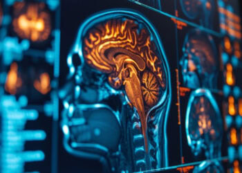
A neuroimaging study comparing individuals with insomnia to healthy adults found disrupted connectivity in the sensory processing cortex and subcortical nuclei of the brain among those with insomnia. This dysfunction likely contributes to the imbalance in their sleep-wake cycles. The findings were published in Brain Imaging and Behavior.
Insomnia is a common sleep disorder that makes it difficult to fall asleep, stay asleep, or return to sleep after waking up too early. It can be short-term (acute) or long-term (chronic), lasting from a few days to several months or more. People with insomnia often feel tired during the day, which can negatively affect concentration, mood, and performance.
Many factors can contribute to insomnia, including stress, anxiety, depression, poor sleep habits, and medical conditions. Lifestyle factors such as caffeine intake, screen use before bed, and irregular sleep schedules can also play a role. Treatment typically involves improving sleep hygiene, undergoing cognitive behavioral therapy, or using medication in some cases. Although insomnia can occur at any age, it is more common among older adults and women.
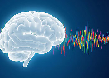
Lead author Hui Wang and colleagues aimed to explore how insomnia develops and identify neural changes that may underlie the disorder using neuroimaging techniques. To do this, they examined a measure called dynamic degree centrality. This metric captures how often and how strongly a specific brain region connects with other regions over time. Unlike static measures, dynamic degree centrality reflects moment-to-moment changes in a region’s connectivity, indicating its shifting importance within brain networks during different mental states or tasks.
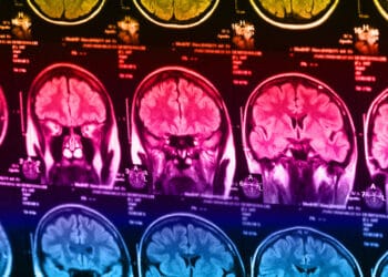
The study involved 29 individuals diagnosed with insomnia and 28 healthy control participants. The control group was matched to the insomnia group by gender, age, and education level. All participants underwent magnetic resonance imaging of the brain. The researchers then calculated both static and dynamic degree centrality values from the imaging data. Participants also completed assessments of sleep quality using the Pittsburgh Sleep Quality Index and levels of depression using the Hamilton Depression Scale.
The results revealed that individuals with insomnia had higher static degree centrality in brain areas associated with sensory processing, such as the occipital gyrus, inferior temporal gyrus, and supramarginal gyrus. Conversely, they showed lower static degree centrality in regions including the parahippocampal gyrus, amygdala, insula, and thalamus—areas involved in emotional processing, memory, and sensory integration.
The researchers also observed group differences in dynamic degree centrality in several regions, including the parahippocampal gyrus, anterior cingulate cortex, medial superior frontal gyrus, inferior parietal gyrus, and precuneus. Notably, higher dynamic degree centrality in the inferior parietal gyrus was associated with better sleep quality. On the other hand, higher static degree centrality in the inferior temporal gyrus correlated with more severe depressive symptoms.
“These findings suggest that dysfunction in centrality within the sensory processing cortex and subcortical nuclei may be associated with the sleep–wake imbalance in individuals with insomnia disorder, contributing to our understanding of hyperarousal mechanisms in insomnia,” the study authors wrote.
The study adds to the scientific knowledge about neural correlates of sleep disorders. However, it should be noted that the design of the study does not allow any causal inferences to be derived from the results. Additionally, the study was conducted on a very small group of participants. Results on larger groups of participants might not be identical.
The paper, “Abnormal sensory processing cortex in insomnia disorder: a degree centrality study,” was authored by Hui Wang, Haining Li, Ziyi Liu, Chiyin Li, Zhaoyao Luo, Wei Chen, Meiling Shang, Huiping Liu, Fatemeh Naderi Nejad, Yuanping Zhou, Ming Zhang, and Yingxiang Sun.


