![[Adobe Stock]](https://sp-ao.shortpixel.ai/client/to_webp,q_glossy,ret_img,w_750,h_375/https://www.psypost.org/wp-content/uploads/2024/10/pinael-gland-thalamus-750x375.jpg)

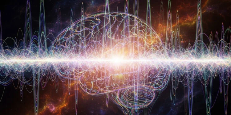
A new study published in Science has identified the thalamus as a central player in how humans become consciously aware of visual information. By recording electrical activity directly from the brains of five patients during a visual task, scientists discovered that specific thalamic regions activate earlier and more strongly during moments of visual awareness. These findings suggest that the intralaminar and medial thalamic nuclei act as a gateway that initiates conscious perception by influencing the activity of the prefrontal cortex.
The thalamus is a small, egg-shaped structure located deep in the center of the brain. It acts as a hub for relaying sensory information—like sight, sound, and touch—to the cerebral cortex, where perception and interpretation occur. In addition to this relay role, it also helps regulate alertness, sleep, and attention.
While the thalamus has long been understood as a relay station for sensory information, it has been largely viewed as a supporting actor—important for maintaining wakefulness, but not the actual source of conscious experience. The current study challenges this idea, offering new evidence that the thalamus does more than just relay signals. It may actively shape what enters our conscious awareness.
To investigate this, the research team took advantage of a unique opportunity to record deep brain activity in humans. Five adult men undergoing treatment for severe, drug-resistant headaches had stereoelectroencephalography (sEEG) electrodes implanted in their brains. These electrodes allowed researchers to monitor neural activity in real-time across multiple brain regions, including various parts of the thalamus and the prefrontal cortex.
Participants completed a visual awareness task that presented images at the edge of conscious visibility. On each trial, a patterned stimulus briefly appeared to one side of a central fixation point. Some images were bright and clearly visible, while others were faint and difficult to detect. Occasionally, no stimulus was shown at all. After each trial, participants responded with an eye movement indicating whether they had seen the stimulus and where it had appeared.
This setup allowed the researchers to compare brain activity between trials where participants consciously saw the stimulus and trials where they did not, even though the visual input was nearly identical. This critical contrast helped isolate the neural processes specifically linked to awareness, rather than simple sensory input.
The results showed that certain regions deep within the thalamus—specifically the central medial nucleus, mediodorsal medial nucleus, and parafascicular nucleus—showed increased activity during trials when participants reported seeing the stimulus. These regions are known as the intralaminar and medial thalamic nuclei. Their responses occurred earlier and with greater intensity than activity in other thalamic regions, such as the ventral nuclei, and even earlier than responses in the prefrontal cortex.
Using a range of analyses, the researchers found that these high-order thalamic nuclei exhibited stronger electrical signals, increased low-frequency oscillations, and more synchronized activity during moments of awareness. Importantly, the timing of this activity—beginning around 200 milliseconds after stimulus presentation—aligned with the earliest known brain signatures of conscious perception, such as the visual awareness negativity signal recorded in prior scalp-based studies.
To understand how these signals travel through the brain, the researchers examined how different brain regions communicated during conscious versus unconscious trials. They measured the synchronization of neural oscillations, particularly in the theta frequency range (about 4–8 Hz), which has been linked to cognitive control and awareness. They also analyzed cross-frequency coupling, which describes how slow brain rhythms may coordinate faster activity in other areas. Both measures revealed that the intralaminar and medial thalamic regions drove neural coordination with the prefrontal cortex during conscious perception.
In other words, when participants were aware of the stimulus, the thalamus was not simply reacting to sensory input—it was actively shaping the brain’s response, sending signals to the prefrontal cortex and helping to organize brain-wide activity in support of awareness.
Additional analyses showed that the neural patterns in the thalamus and prefrontal cortex encoded information about whether participants were consciously aware of the stimulus, rather than simply reflecting aspects of the task like contrast strength, motor response, or reaction time. This strengthens the case that these regions are directly involved in conscious perception, rather than just downstream reflections of other cognitive processes.
Interestingly, the researchers also found that the activity in the thalamus, particularly in the intralaminar and medial nuclei, could begin even before the stimulus appeared. This pre-stimulus activity was more synchronized in trials where participants would later report being aware of the stimulus, suggesting that these regions may help set the stage for conscious perception by influencing the brain’s state of readiness.
This study is the first to offer such detailed recordings from multiple thalamic nuclei in humans during a task designed to probe the boundary between conscious and unconscious visual perception. Prior studies using imaging methods like functional MRI or magnetoencephalography hinted at thalamic involvement in awareness but lacked the precision to pinpoint when and where this activity occurred. The use of sEEG here provided a rare opportunity to capture the dynamics of deep brain structures with both high temporal and spatial resolution.
The findings provide strong support for the idea that consciousness is not solely a cortical phenomenon. Instead, the thalamus—especially its high-order nuclei—appears to play an active and early role in making sensory information available to our awareness. This may reflect the thalamus’s unique position as a hub, with widespread connections to both cortical and subcortical brain regions, allowing it to coordinate complex patterns of neural activity required for consciousness to emerge.
However, the authors note some limitations. The study was conducted in a small sample of five male patients, all of whom had electrodes implanted for clinical reasons related to headache treatment. Although their cognitive abilities and vision were intact, these participants do not represent the general population. Moreover, electrode placement was determined by medical need, not research goals, which means some brain regions may have been under-sampled or missed entirely.
Additionally, while the study strongly suggests that intralaminar and medial thalamic nuclei initiate the conscious perception process, it does not rule out important contributions from the cortex. The prefrontal cortex, particularly the lateral regions, still showed significant activity and participated in synchronized neural networks during awareness. The researchers suggest that the thalamus may act as a gate or initiator, while the cortex elaborates on the content of experience.
The study, “Human high-order thalamic nuclei gate conscious perception through the thalamofrontal loop,” was authored by Zepeng Fang, Yuanyuan Dang, An’an Ping, Chenyu Wang, Qianchuan Zhao, Hulin Zhao, Xiaoli Li, and Mingsha Zhang.


