
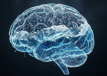
A new study published in iScience provides evidence that the human brain emits extremely faint light signals that not only pass through the skull but also appear to change in response to mental states. Researchers found that these ultraweak light emissions could be recorded in complete darkness, and they appeared to shift in response to simple tasks like closing the eyes or listening to sound. The findings suggest that this faint brain light may carry information about brain activity—possibly opening the door to a new way of studying the brain (photoencephalography).
All living tissues release tiny amounts of light during normal metabolism, known as ultraweak photon emissions. This happens when excited molecules return to a lower energy state and emit a photon in the process. The light is incredibly faint—about a million times weaker than what we can see—and falls within the visible to near-infrared range. In contrast to bioluminescence, which involves specific chemical reactions like those used by fireflies, ultraweak photon emissions happen constantly in all tissues, without special enzymes or glowing compounds.
The brain emits more of this faint light than most other organs because of its high energy use and dense concentration of photoactive molecules. These include compounds like flavins, serotonin, and proteins that can absorb and emit light. Photon emission rates also seem to rise during oxidative stress and aging and may reflect changes in cell health or communication.
The research team, led by Hayley Casey, Nirosha Murugan, and colleagues at Algoma University, Tufts University, and Wilfrid Laurier University, wanted to know if these faint light emissions could be used to monitor brain activity. Unlike other imaging methods that require stimulation—such as strong magnetic fields or infrared light—measuring UPEs is entirely passive. That means it doesn’t introduce anything new to the brain.
The researchers proposed that UPEs might offer a new way to monitor brain function safely and without interference, similar to how EEG tracks electrical brain waves without applying energy. They also wanted to test whether UPEs reflect mental states like resting with eyes closed or responding to sound, and whether these signals match known changes in electrical brain rhythms.
The researchers recruited 20 healthy adult participants and measured both UPEs and brain electrical activity while the participants sat in a dark room. The setup included photomultiplier tubes placed near the occipital and temporal regions of the head, where the brain processes visual and auditory information. A third sensor recorded background light. At the same time, participants wore a cap with electroencephalography sensors to record electrical brain rhythms.
Participants went through a ten-minute recording session that included five conditions. First, they sat with eyes open and then with eyes closed. Next, they listened to a simple repeating auditory stimulus, followed by another eyes-closed period, and finally another eyes-open period. The aim was to see whether brain UPEs responded to known manipulations of brain activity, particularly the shift in alpha rhythms that occurs when people close their eyes.
Photon emissions were recorded in short time intervals and analyzed for variability, frequency content, and stability over time. The team compared the results to background signals and examined correlations with electrical brain rhythms recorded at the same time.
The researchers found that the light emitted from the brain could be distinguished from background light based on its variability and complexity. Brain UPEs showed greater entropy and a more dynamic signal than background recordings. These emissions also showed a distinctive frequency profile below 1 Hz, meaning that the light fluctuated in slow rhythmic patterns roughly once every one to ten seconds. This signature was not present in the background light and was especially pronounced in the occipital region.
The researchers also observed that brain UPEs appeared to reach a steady state during each task, particularly by the end of the two-minute recording segments. These stable patterns shifted when participants moved between eyes-open and eyes-closed conditions, suggesting that the emissions reflect changes in the brain’s internal state. However, the direction of change was not consistent across all participants, possibly reflecting individual differences or the complexity of the underlying metabolic processes.
When the researchers compared UPEs to brain electrical rhythms, they found modest correlations. For instance, alpha rhythms—often associated with relaxed wakefulness and increased during eyes-closed states—correlated with photon emissions from the occipital region, but only when the participants’ eyes were closed. They also found some associations between UPE variability and temporal lobe rhythms during auditory stimulation. Still, the relationships were not strong, and many expected correlations did not emerge, highlighting the need for further research.
While the findings are promising, the authors caution that this was a preliminary study with several limitations. The sample size was small, and the recording equipment only covered a few areas of the head. The sensors also detected a wide range of wavelengths, which may have obscured more specific light patterns. More precise filters or detectors could help uncover wavelength-specific signatures related to different brain functions.
The researchers also suggest that expanding the sensor array could improve spatial resolution and help identify the origins of UPEs within the brain. Because these emissions are tied to metabolic activity, they may come from a variety of cell types, including neurons and glial cells, and from different depths in the brain. Developing methods to localize these signals will be an important next step.
The study did not include measurements from other parts of the body, which could help clarify whether similar light emissions occur in non-brain tissues and how they differ. Including more participants and exploring variations based on age, sex, or health status may also reveal meaningful patterns. In the future, machine learning and advanced imaging techniques may allow researchers to decode UPE patterns and use them to detect brain disorders or monitor brain health.
“We view the current results as a proof-of-concept demonstration that patterns of human-brain-derived UPE signals can be discriminated from background light signals in darkened settings despite very low relative signal intensity,” the researchers wrote. “Photoencephalography would be maximally non-invasive (i.e., passive recording) with high temporal resolution, like EEG or MEG; however, the measurement of UPEs would be linked to oxidative metabolism with several clinical applications described elsewhere. Future studies may find success in using select filters and amplifiers to sieve and enhance UPE signal features from healthy and diseased brains.”
The study, “Exploring ultraweak photon emissions as optical markers of brain activity,” was authored by Hayley Casey, Isabella DiBerardino, Mattia Bonzanni, Nicolas Rouleau, and Nirosha J. Murugan.


