

Substance use disorders, which include addiction to substances like alcohol, nicotine, cocaine, opioids, and cannabis, are complex and challenging conditions that affect millions of people worldwide. Understanding the underlying brain mechanisms of these disorders is crucial for effective treatment and intervention. A recent study, published in Nature Mental Health, sheds light on the common brain network that may be at the heart of various substance use disorders, providing valuable insights for future treatment approaches.
Substance use disorders pose a significant public health concern, contributing to disability, mortality, and substantial economic costs. In the United States alone, over 20 million individuals grapple with substance use disorders, resulting in annual costs exceeding $700 billion.
Neuroimaging studies play a vital role in understanding these disorders, but the existing literature is characterized by variability in imaging methods and analysis approaches. Previous studies often focused on specific substances, making it challenging to identify commonalities between different substance use disorders.
Traditionally, researchers have attempted to pinpoint brain regions associated with addiction to particular substances. However, this approach has yielded mixed and inconsistent results. Meta-analyses, which combine findings from multiple studies, have provided varying evidence for convergent neuroimaging abnormalities but have not consistently implicated the same brain regions across different substances or imaging modalities.
“There have been many studies that have looked at neuroimaging in substance use disorder and addiction. We were primarily interested in doing this work because previous studies implicated many different brain regions and so the underlying circuitry, and potential therapeutic targets, remained unclear,” said study author Jacob Stubbs, who completed this study while at Brigham and Women’s Hospital, working with Michael Fox and the team at the Center for Brain Circuit Therapeutics. He is currently a medical student at the University of British Columbia.
In response to these challenges, the study introduced a novel approach called ‘coordinate network mapping’ to investigate whether heterogeneous neuroimaging abnormalities in substance use disorders are linked to a common brain network. This approach had previously been successful in mapping common networks in other diseases and disorders, offering a promising avenue for understanding addiction better.
To unravel the common brain network associated with substance use disorders, the researchers analyzed data from 144 imaging studies involving more than 9,000 participants. These studies covered a wide range of substances and employed various imaging techniques, making it a comprehensive investigation into the underlying neural mechanisms.
The researchers first focused on 45 studies that examined brain atrophy related to substance use disorders. Atrophy refers to the loss of neurons and their connections in the brain, often seen in individuals with chronic substance abuse. This analysis included data from nearly 3,800 participants addicted to substances like alcohol, nicotine, cocaine, opioids, and cannabis.
Next, the researchers analyzed data from 99 studies that used functional magnetic resonance imaging (fMRI) to assess over 5,000 individuals with substance use disorders. This step aimed to understand the brain’s functional abnormalities associated with addiction, irrespective of the specific substance involved.
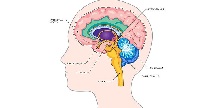
The researchers found that a remarkable 91% of the atrophy coordinates from the first set of studies mapped to a common brain network. This network included regions like the anterior cingulate, insulae, prefrontal cortices, and the thalamus. These areas are known to be involved in processes related to addiction, such as craving, emotion, and risky decision-making. Importantly, this network was distinct from the brain regions affected by normal aging and neurodegenerative diseases.
Moving on to the fMRI data, the researchers discovered that over 80% of the neuroimaging abnormalities associated with substance use disorders mapped to a similar brain network, regardless of the specific substance involved. This finding suggests that there is a common neural circuitry underlying addiction, regardless of the substance being abused.
The study’s combined analysis across different substances and imaging methods revealed a consistent common brain network. This discovery is significant because it provides researchers and clinicians with a specific target for interventions and treatments, rather than focusing on isolated brain regions.
“We found that brain imaging abnormalities in substance use disorder and addiction are connected to a common brain circuit,” Stubbs told PsyPost. “This helps improve our understanding of the brain circuits linked to addiction and provides a brain network that we might be able to target for better therapies. ”
While this study provides critical insights into the common brain network underlying substance use disorders, it is essential to acknowledge some limitations. The data used in the study encompassed diverse imaging approaches, software packages, and demographic variables across different studies. This heterogeneity could introduce noise into the findings. Additionally, the study focused on identifying common networks, and future research should explore substance-specific networks in more detail, potentially using larger sample sizes or more granular data.
Despite these limitations, the alignment of coordinate and lesion networks in this study offers valuable insights into the therapeutic potential of targeting the identified brain network. Transcranial magnetic stimulation, an FDA-approved treatment for nicotine use disorder, has targeted regions within this network, suggesting promising avenues for addiction treatment.
The study, “Heterogeneous neuroimaging findings across substance use disorders localize to a common brain network“, was authored by Jacob L. Stubbs, Joseph J. Taylor, Shan H. Siddiqi, Frederic L. W. V. J. Schaper, Alexander L. Cohen, William Drew, Colleen A. Hanlon, Amir Abdolahi, Henry Z. Wang, William G. Honer, William J. Panenka, and Michael D. Fox.


