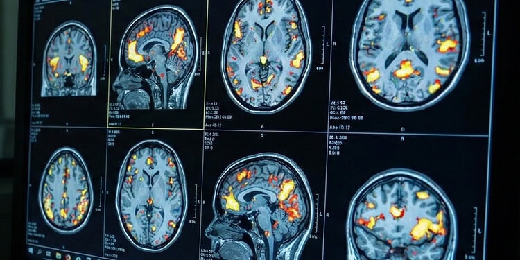

A small study suggests that the primary method used to map brain activity may often yield reversed metabolic readings. The research reveals that blood oxygen levels do not consistently reflect underlying energy consumption in the brain, challenging standard interpretations of brain imaging. The findings were published in Nature Neuroscience.
Functional magnetic resonance imaging, or fMRI, is a standard tool for observing the brain in action. When people perform tasks inside an MRI scanner, the machine does not actually see electrical impulses from neurons. Instead, it detects changes in blood oxygen levels across different brain regions.
This measurement is known as the blood-oxygenation-level-dependent signal, or BOLD signal. The conventional interpretation assumes that an increase in the BOLD signal reflects a surge in local brain activity.
Neurons require energy to function, and they obtain this energy by consuming oxygen. Because neurons lack their own energy reserves, active brain areas rely on a rapid influx of fresh, oxygen-rich blood. This physiological process usually supplies more oxygen than the neurons immediately consume, which flushes out deoxygenated blood and creates a positive BOLD signal on the brain scan.
Conversely, a drop in the BOLD signal is typically interpreted as a decrease in brain cell activity. However, anomalies in this relationship occasionally surface in animal research and specialized human brain scans. These anomalies suggest that the link between blood flow and oxygen consumption might not be uniform across the entire brain.
More than two decades ago, scientists developed a theoretical framework to predict how the BOLD signal should behave based on varying levels of blood flow and oxygen consumption. This framework suggested that certain biological conditions could produce a BOLD signal that contradicts the actual energy use of the brain cells. Until now, this theory had not been comprehensively tested across the entire human cerebral cortex.
Researchers Samira M. Epp, Valentin Riedl, and colleagues sought to test whether this standard biological assumption holds true across the entire human brain. They designed a study to directly measure oxygen consumption alongside the standard BOLD signal. Epp is a researcher at the Technical University of Munich and the University of Erlangen-Nuremberg, where Riedl also holds an affiliation.
The investigators conducted a small study involving 40 healthy participants. They asked the volunteers to perform a series of mental exercises while inside an MRI machine. The researchers utilized a specialized imaging protocol called quantitative fMRI. This advanced technique captures the standard BOLD signal while also measuring absolute blood flow, blood volume, and the rate of oxygen extraction from the bloodstream.
The scanning session required participants to engage in an arithmetic calculation task. This specific math task was chosen because prior research shows it reliably stimulates attention-related brain networks while simultaneously suppressing activity in the brain’s default mode network. Participants also completed an autobiographical memory task designed to stimulate the default mode network, as well as a basic visual control task to serve as a baseline.
By comparing the imaging data across these varying mental states, the researchers could track both the BOLD signal and the actual amount of oxygen the brain tissue was metabolizing. To ensure their results were robust, they conducted multiple control analyses. These controls included a replication study with ten participants using matched image resolutions to rule out potential scan distortion artifacts.
The results contradicted the standard biological model in a substantial portion of the brain. When the researchers examined areas showing a positive BOLD signal, they found that the canonical model held true on average. Blood flow increased heavily, supplying an excess of oxygen and generating the expected brain scan signature.
However, the results for areas displaying a negative BOLD signal were unexpected. Based on conventional wisdom, a negative signal should indicate that neurons are resting and metabolizing less oxygen. Instead, the researchers found that oxygen consumption did not decrease in these areas, and in some cases, it actually increased despite the negative BOLD reading.
When examining individual volume units of the brain scan, known as voxels, the discrepancies became even more pronounced. A voxel is a three-dimensional pixel representing a tiny cube of brain tissue. The researchers discovered that in about 40 percent of the voxels displaying a strong BOLD response, the actual oxygen metabolism moved in the opposite direction of the BOLD signal. The team labeled these specific brain units as discordant voxels.
In discordant voxels showing a positive BOLD signal, the brain tissue was actually consuming less oxygen than at rest. In discordant voxels showing a negative BOLD signal, the brain tissue was consuming more oxygen. This means that simply looking at the BOLD signal would lead to a backward interpretation of the underlying cellular energy use in nearly half of the responding brain regions.
The team also identified voxels that exhibited a mixed response. In these areas, the tissue demonstrated a standard, expected blood flow response during one cognitive task but shifted to a discordant response during another task. This flexibility suggests that a single region of the brain can alter its oxygen delivery mechanism depending on the specific cognitive demands of the moment.
The researchers identified two distinct biological mechanisms driving these brain responses. In standard, concordant brain regions, the tissue met its energy needs by triggering an immense increase in local blood flow. This massive rush of blood overcompensates for the oxygen consumed by the neurons, generating the classic positive BOLD signal used in most neuroscience studies.
In discordant regions, the tissue handled its energy needs differently. Instead of relying on a large influx of fresh blood, the neurons simply extracted a higher percentage of oxygen from the blood that was already present in the local capillaries. Because the overall blood flow barely changed, this increased oxygen extraction left a higher concentration of deoxygenated blood in the area.
Deoxygenated blood has magnetic properties that darken the MRI image. As a result, this higher extraction rate caused a negative BOLD signal, even though the neurons were actively working and consuming more energy.
The researchers noticed that these two types of brain tissue function differently even when a person is resting. Discordant regions naturally maintain a lower baseline rate of oxygen extraction. This lower baseline provides them with a built-in reserve of oxygen to draw from when they suddenly need to perform a cognitive task, allowing them to function without demanding a fresh surge of blood from the vascular system.
One limitation of quantitative MRI techniques is that measuring total blood volume can be technically challenging. The method used in the study captures total blood volume rather than strictly venous volume, which can introduce some variation when comparing data between subjects. Additionally, the calculations for oxygen metabolism are primarily validated for the brain’s outer gray matter and are difficult to interpret in the deep white matter.
Readers should not interpret these results as invalidating decades of prior fMRI research. A positive BOLD signal still reliably indicates increased oxygen metabolism on a broad, average scale. However, the exact location and magnitude of that activity may be misinterpreted if scientists rely exclusively on standard BOLD imaging without considering regional differences in blood flow.
Future imaging studies might need to adopt quantitative fMRI methods to accurately map brain function. This approach could be especially relevant for studying aging populations or patients with vascular diseases, where blood flow responses might be altered. Tracking actual oxygen consumption alongside standard imaging provides a more accurate window into how the human brain distributes its energy.
The study, “BOLD signal changes can oppose oxygen metabolism across the human cortex,” was authored by Samira M. Epp, Gabriel Castrillón, Beijia Yuan, Jessica Andrews-Hanna, Christine Preibisch, and Valentin Riedl.