![[Adobe Stock]](https://sp-ao.shortpixel.ai/client/to_webp,q_glossy,ret_img,w_750,h_375/https://www.psypost.org/wp-content/uploads/2024/11/brain-scan-750x375.jpg)
Researchers have identified specific coupled patterns of brain activity and gene expression that help explain impulsive behavior in children with attention deficit hyperactivity disorder. By analyzing these two distinct types of biological data together, the study demonstrates that the combination predicts behavioral symptoms more accurately than looking at either brain scans or genetic information in isolation. These findings were published in the journal Biological Psychiatry: Cognitive Neuroscience and Neuroimaging.
Attention deficit hyperactivity disorder is a neurodevelopmental condition that affects a large portion of the global childhood population. It is often characterized by difficulty paying attention, excessive activity, and impulsivity. Impulsivity involves acting without foresight or inhibiting one’s immediate reactions. This specific symptom can lead to challenges in social settings and academic performance.
Researchers have known for some time that this disorder is highly heritable. Estimates suggest that genetics account for 70 to 80 percent of the risk for developing the condition. Despite this strong hereditary link, scientists have struggled to pinpoint specific genetic variants that account for a large portion of the disorder. Genome-wide association studies often identify genetic variations with very small individual effects.
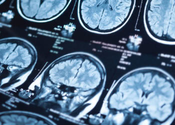
Simultaneously, neuroscientists use tools like functional magnetic resonance imaging to study the brain in action. These scans measure blood flow changes to determine which parts of the brain are working during specific tasks. Previous imaging research has shown that children with the disorder often exhibit distinct activity patterns in brain networks responsible for attention and inhibition. However, studies rarely combine these functional brain maps with detailed genetic data in a single analysis.
To address this gap, a team of researchers led by Soohyun Jeon and Jong-Hwan Lee from the Department of Brain and Cognitive Engineering at Korea University set out to integrate these two modalities. They aimed to find a hidden relationship between the brain’s functional activity and the expression of specific gene sets. They hypothesized that combining these data points would create a stronger model for predicting impulsive behavior.
The research team utilized data from the Adolescent Brain Cognitive Development study. This is a massive, long-term project tracking the biological and behavioral development of children across the United States. For this specific investigation, the authors selected 394 children diagnosed with attention deficit hyperactivity disorder and 1,000 healthy control subjects. The participants were all between the ages of nine and ten. To ensure genetic homogeneity for this specific analysis, the team focused on children of European ancestry.
The researchers assessed impulsivity using a computerized test known as the stop signal task. In this activity, children are told to press a button when they see a “Go” signal. Occasionally, a “Stop” signal appears shortly after the first prompt. The child must then inhibit their impulse to press the button. The time it takes for a child to successfully stop an initiated response provides a quantitative measure of their motor impulsivity.
While the children performed this task, an MRI machine recorded their brain activity. In addition to the imaging data, the researchers analyzed the participants’ genetic information. Instead of looking only at raw genetic code, they used a computational method to estimate gene expression levels in specific brain tissues. This approach predicts how active certain genes are likely to be in different parts of the brain based on the child’s genetic profile.
To analyze this complex set of information, the team employed a sophisticated statistical technique called parallel independent component analysis. This method allows scientists to process two different types of data simultaneously. It searches for components or patterns in the brain scan data that statistically correlate with components in the gene expression data.
The researchers split the data into two groups to ensure their results were robust. They used 80 percent of the participants as a “discovery” set to find the patterns. They reserved the remaining 20 percent as a “replication” set to test if those patterns held true in a fresh batch of data. This step is essential for validating scientific results and ensuring they are not merely random chance.
The analysis revealed three distinct pairs of linked brain and gene patterns. These pairs appeared consistently in the cortex, the cerebellum, and the nucleus accumbens. Each pair represented a connection between activity in that specific brain region and the expression of a specific group of genes.
The first pair involved the cerebral cortex. The brain scans showed activity in areas responsible for decision-making and stopping movements, such as the orbitofrontal gyrus and the medial prefrontal gyrus. The associated genetic component included genes related to immune function and metabolism. This aligns with emerging theories that inflammation and immune system regulation may play a role in psychiatric conditions.
The second pair centered on the cerebellum. This structure at the back of the brain is traditionally associated with balance and motor control. However, it is increasingly recognized for its role in cognitive processes. The imaging data showed patterns in the default mode network, a set of brain regions active when the mind is at rest but typically suppressed during focused tasks. The genetic counterpart included genes that regulate how other genes function.
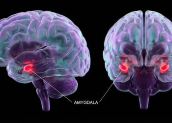
The third pair was linked to the nucleus accumbens. This region is a central part of the brain’s reward system. The analysis highlighted connections between this area and the frontal cortex. The genetic profile associated with this component involved genes linked to inflammation and synaptic plasticity, which is the ability of neurons to strengthen or weaken their connections.
The most practical outcome of the study was the improvement in predicting behavioral symptoms. The researchers tried to predict the children’s scores on the stop signal task using different statistical models. Models that relied solely on the brain scan data or solely on the genetic data provided limited predictive power.
However, when the researchers combined the paired components—using both the brain activity and the gene expression data—the accuracy of the predictions improved. The model became even more sensitive when it accounted for the interaction between the two data types. For example, in predicting the “stop signal reaction time,” which measures how quickly a child can cancel a movement, the combined model showed a substantial boost in performance compared to the single-modality models.
For children with attention deficit hyperactivity disorder, the combined model also improved the prediction of reaction time variability. This metric tracks how consistent a child is when responding to the “Go” signal. High variability is a common trait in the disorder and reflects lapses in attention. The study showed that the interaction between the cortical brain patterns and the associated gene expressions helped explain this inconsistency.
The study also found a connection to intelligence scores. In the group of children with the disorder, the linked pattern in the cerebellum was highly predictive of IQ. This relationship was less apparent in the healthy control group. This suggests that the biological factors driving cognitive performance may differ between neurotypical children and those with attention deficits.
There are limitations to this research that require consideration. The study focused exclusively on children of European ancestry. This decision was made to reduce genetic variation that could complicate the analysis, but it means the results may not apply to children of other backgrounds. Future studies must include more diverse populations to ensure the findings are universally applicable.
Another caveat involves the source of the gene expression data. The researchers estimated gene activity based on adult post-mortem tissue databases. Gene expression changes as the brain develops. Consequently, the estimated activity in a ten-year-old’s brain might differ from the adult patterns used as a reference. Future research would benefit from using gene expression databases derived specifically from pediatric tissue.
Additionally, the brain imaging data came from multiple sites across the country using different MRI scanners. While the researchers used statistical methods to correct for this, slight variations in scanner hardware can introduce noise into the data.
Despite these limitations, the study represents a step forward in the field of imaging genetics. It moves beyond looking at single genes or isolated brain regions. Instead, it views the disorder through a multivariate lens, recognizing that biological systems work in concert. The authors emphasize that this “biologically plausible” approach helps bridge the gap between microscopic molecular processes and macroscopic brain behavior.
The study, “Abnormal association between neural activity and genetic expressions of impulsivity in attention deficit hyperactivity disorder: an Adolescent Brain Cognitive Development study,” was authored by Soohyun Jeon, Jae-eon Kang, Jundong Hwang, Vince D. Calhoun, and Jong-Hwan Lee.


