For decades, scientists have sought to understand the biological foundations of genius by examining the physical characteristics of Albert Einstein’s brain. Various studies indicate that while his brain weighed a normal amount, it possessed distinct anatomical features and enhanced connectivity that may have contributed to his extraordinary cognitive abilities. These findings offer a glimpse into the potential neural substrates of his mathematical and visuospatial prowess.
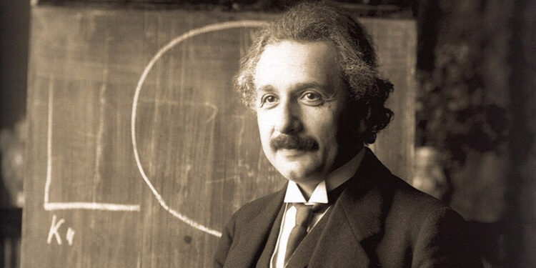
Albert Einstein is widely recognized as one of the most influential physicists in history. In 1905, often referred to as his annus mirabilis or miraculous year, he published four groundbreaking papers that fundamentally altered the scientific understanding of the universe. These works addressed the photoelectric effect, Brownian motion, special relativity, and the equivalence of mass and energy. He later developed the general theory of relativity, cementing his reputation as a scientific revolutionary.
Because of his immense intellectual achievements, significant curiosity surrounded the source of his genius. Researchers and the public alike questioned whether his abilities stemmed from his environment and education or if he possessed a unique biological advantage. This debate regarding nature versus nurture prompted a desire to analyze his physical brain for clues.
Einstein died on April 18, 1955, at Princeton Hospital due to a ruptured abdominal aortic aneurysm. During the autopsy, the pathologist Thomas Harvey removed Einstein’s brain for scientific study. Harvey weighed the organ, photographed it from multiple angles, and preserved it using formalin. He subsequently sectioned the brain into approximately 240 blocks and prepared histological slides, which are microscopic slices of tissue used for studying cellular structure.
A Review of the Research
Despite the early preservation of the tissue, scientific analysis appeared slowly. A review article published in Neurosciences and History in 2015 by Paul Carrillo-Mora and colleagues provides a chronological overview of the major studies conducted on the specimens. The authors note that thirty years passed between Einstein’s death and the first publication of data regarding his brain’s morphology.
The review highlights that researchers have identified multiple microscopic and macroscopic differences between Einstein’s brain and control samples. However, Carrillo-Mora and his team note that the functional significance of these anomalies remains a topic of debate. They suggest that while structural peculiarities exist, determining their direct link to genius requires careful interpretation.
Cellular Composition
One of the first major studies appeared in Experimental Neurology in 1985. Marian C. Diamond and her colleagues at the University of California, Berkeley, examined the ratio of neurons to glial cells in specific regions of the brain. Neurons are the primary cells responsible for transmitting information, while glial cells provide support, nutrients, and insulation to neurons.
The researchers analyzed samples from the prefrontal cortex and the inferior parietal lobes. The prefrontal cortex associates with planning and abstract thought, while the parietal lobes handle sensory integration. Diamond compared Einstein’s tissue to samples from eleven male control subjects.
The study found that in the left posterior parietal lobe, Einstein’s brain possessed a significantly lower ratio of neurons to glial cells. This indicates a higher number of glial cells for every neuron. Diamond suggested that this increased presence of support cells might reflect a higher metabolic need. The neurons in this region of Einstein’s brain may have been utilizing more energy, requiring more glial cells to support their intense activity.
Anatomical Structure and the Parietal Lobes
In 1999, Sandra Witelson and her team at McMaster University published a study in The Lancet that focused on the gross anatomy, or visible structure, of the brain. They utilized photographs taken by Harvey in 1955 and measurements taken directly from the organ. They compared these against a control group of thirty-five male and fifty-six female brains.
Witelson found that Einstein’s brain weight was not exceptional. However, the parietal lobes displayed striking differences. The study revealed that his parietal lobes were approximately 15 percent wider than those of the control group.
Furthermore, the researchers identified a unique pattern in the Sylvian fissure. This fissure is a deep fold that separates the temporal lobe from the frontal and parietal lobes. In typical brains, this fissure curves up and divides a region called the supramarginal gyrus.
In Einstein’s brain, the Sylvian fissure followed an unusual trajectory that left the supramarginal gyrus undivided. Witelson proposed that this absence of division allowed for more efficient connections between neurons in that area. This region supports visuospatial cognition and mathematical thinking. The study suggests this unique anatomy may have facilitated the type of visual thinking Einstein used to develop his theories.
Surface Features and Motor Skills
In 2009, anthropologist Dean Falk published a study in Frontiers in Evolutionary Neuroscience that applied techniques from paleoanthropology to analyze photographs of Einstein’s brain. This field usually focuses on the evolution of human anatomy, but Falk used these skills to identify previously overlooked details on the brain’s surface.
Falk identified an unusual “knob-like” structure on the right motor cortex. The motor cortex is the area of the brain that controls voluntary muscle movement. This specific knob corresponds to the area that controls the left hand.
Falk noted that this feature is often seen in long-time musicians. Einstein famously played the violin from childhood. The study implies that this anatomical feature developed as a result of the intense, repetitive motion required to finger the strings with his left hand. This finding provides evidence for plasticity, the brain’s ability to physically change in response to repeated activity.
Falk also observed unusual grooving patterns in the parietal lobes. These patterns reinforced earlier findings regarding the expansion of association cortices. These are areas responsible for integrating information from different senses.
Interhemispheric Connectivity
A study published in the journal Brain in 2013 provided further insight into how the two halves of Einstein’s brain communicated. Weiwei Men, Dean Falk, and colleagues analyzed the corpus callosum. This structure is a thick bundle of nerve fibers that acts as a bridge connecting the left and right cerebral hemispheres.
The researchers used high-resolution photographs to measure the thickness of the corpus callosum at various points. They compared these measurements to MRI data from two control groups: fifteen elderly men and fifty-two young men.
The analysis revealed that Einstein’s corpus callosum was thicker than that of the controls in most subregions. A thicker corpus callosum implies a higher number of nerve fibers. This suggests that Einstein possessed enhanced connectivity between the two hemispheres of his brain.
Increased connectivity allows for faster and more integrated processing of information. The authors proposed that this facilitated cooperation between the analytical functions often associated with the left hemisphere and the spatial or creative functions of the right hemisphere. This neural integration may have supported his ability to visualize complex physics problems.
Rediscovering Lost History
Much of the recent understanding of Einstein’s brain comes from the recovery of materials thought to be lost. In his book Finding Einstein’s Brain, Frederick Lepore details the history of these biological specimens. Lepore, a neurologist at Robert Wood Johnson Medical School, collaborated with Dean Falk to analyze photographs that had remained in the possession of Thomas Harvey’s family for decades.
Following Harvey’s death in 2007, his family donated the materials to the National Museum of Health and Medicine. This collection included dozens of photographs and hundreds of histological slides. Lepore explains that these materials provided a view of the brain before it was dissected, allowing for a more comprehensive analysis of its surface anatomy.
The book and associated research highlighted a significant anomaly in the frontal lobe. The frontal lobe is the part of the brain located directly behind the forehead. It manages executive functions, such as planning, working memory, and decision-making.
In a typical human brain, the frontal lobe contains three ridges, known as gyri. Lepore and Falk identified that Einstein’s right frontal lobe possessed a fourth gyrus. This extra ridge suggests an increase in the surface area of the prefrontal cortex.
This region associates closely with high-order cognitive processes. The presence of additional cortical surface area implies a greater capacity for the complex mental simulations Einstein employed. He famously used “thought experiments,” such as imagining chasing a beam of light, to conceptualize his theories.
The Parietal Lobe Revisited
Lepore’s work also reinforces the significance of the parietal lobe asymmetry. The book details how the inferior and superior parietal lobules were grossly asymmetrical. This contradicts earlier assumptions that Einstein’s brain was spherical.
The text explains that the parietal lobes are essential for processing sensory information and spatial orientation. The unusual expansion in these areas aligns with reports of Einstein’s preference for thinking in images rather than words. He often described his thought process as a combinatory play of images and signs.
Skepticism and Context
It is important to note that some members of the scientific community remain skeptical of these findings. Critics argue that attributing Einstein’s genius to specific anatomical bumps or ridges resembles phrenology, a discredited pseudoscience. They contend that natural human variation is vast.
Lepore addresses this in his writings by emphasizing that the research presents data points rather than definitive explanations. He argues that while one cannot bridge the explanatory gap between brain structure and the mind entirely, the anatomical deviations are too significant to ignore. He posits that the unique structure provides the hardware that made his cognitive software possible.
Conclusion
The study of Albert Einstein’s brain offers a fascinating intersection of history, neuroscience, and biography. Research indicates that his genius was likely not the result of a single anomaly but rather a combination of features. These include a higher ratio of glial cells, expanded parietal lobes, a unique ridge in the frontal cortex, and enhanced connectivity between hemispheres.
These biological traits appear to correlate with the specific cognitive strengths Einstein displayed, particularly his mathematical insight and visual thinking. While the mystery of genius cannot be solved by anatomy alone, the evidence suggests that Einstein’s brain was physically adapted for the unique intellectual tasks he performed. The recovered photographs and slides continue to provide a resource for scientists seeking to understand the neural machinery of a brilliant mind.


