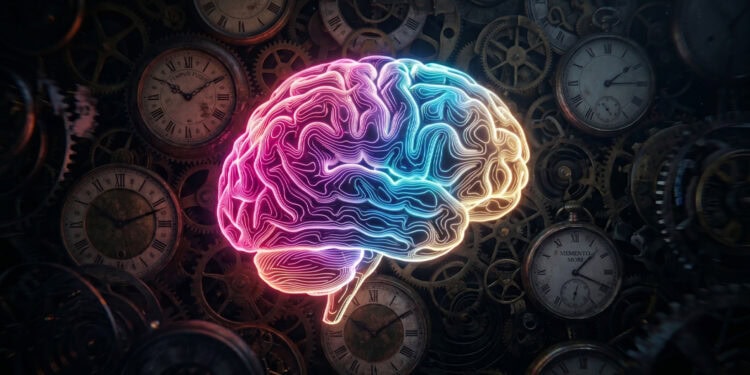
A recent study mapping the human brain reveals that our perception of time does not happen all at once, but rather unfolds across a series of distinct physical processing stages. As visual information travels from the back of the brain to the front, different groups of neurons handle specific parts of the timing process, ultimately creating our subjective experience of how long an event lasts. These findings were published in the journal PLOS Biology.
For decades, researchers have mapped out a broad network of brain regions that become active when people estimate how much time has passed. Studies involving both animals and humans have shown that certain groups of neurons respond to specific durations of time.
These specialized cells are often arranged in topographic maps across the brain. In these maps, neurons that prefer similar lengths of time are located physically close to one another on the folded outer layer of the brain, known as the cerebral cortex.
Despite knowing where these timing regions are located, researchers have struggled to understand exactly how they work together. It has been unclear how a physical feature like the duration of a flashing light is transformed into an abstract feeling of passing time.
To piece together this puzzle, neuroscientist Valeria Centanino and her colleagues Gianfranco Fortunato and Domenica Bueti at the International School for Advanced Studies in Italy conducted an imaging study. They wanted to track how the properties of time-tracking neurons change as signals move through the brain.
The researchers recruited thirteen healthy volunteers to perform a visual categorization task. First, the participants were trained to memorize a specific reference duration of half a second, which they would use as a mental benchmark.
During the main experiment, the volunteers watched a series of blurry, flickering circles appear on a screen. Each circle stayed on the screen for a random amount of time, ranging between two-tenths of a second and eight-tenths of a second.
After each circle disappeared, the participants pressed a button to indicate whether the shape was visible for a longer or shorter time than their internalized reference. While the volunteers performed this task, the researchers recorded their brain activity using an ultra-high-field functional magnetic resonance imaging scanner.
Functional magnetic resonance imaging is a technology that measures brain activity by detecting changes in blood flow. When a specific area of the brain works harder, it requires more oxygen, and the scanner tracks the oxygen-rich blood rushing to that region.
The scanner used in this study operates at a magnetic field strength of seven Tesla. This is much stronger than standard hospital scanners, allowing the team to capture highly detailed images of the brain surface.
With these detailed images, Centanino and her team modeled the behavior of individual populations of neurons. They looked for unimodal tuning, which happens when a group of brain cells responds most strongly to one specific stimulus and less strongly to anything else.
The researchers found that the way neurons tuned into time changed depending on their location in the brain. They identified three distinct processing stages that form a hierarchy of time perception.
The first stage occurs in the occipital visual areas, located at the back of the head where the brain first processes sight. Here, the neurons acted like simple timers that gathered sensory information from the eyes.
In these visual areas, the brain cells showed a strong preference for the longest durations. Their activity increased steadily the longer the shape stayed on the screen, encoding the physical length of the visual event.
The second stage takes place in the parietal and premotor regions, which sit near the top and middle of the brain. In these areas, the researchers observed a complete topographic map of time.
Neurons in these middle regions were tuned to the entire range of presented durations. Some groups of cells responded only to brief flashes, while others responded only to medium or long appearances.
These specialized cells were neatly organized into clusters based on their preferred durations. This suggests that the parietal and premotor regions are responsible for reading out the specific duration of the visual event, allowing the brain to track exactly how much time just passed.
The final stage happens in the frontal regions of the brain, including the anterior insula and the rostral supplementary motor area. These areas are heavily involved in complex thought, decision making, and self-awareness.
In these frontal areas, the neurons did not represent the full range of time. Instead, they showed a strong preference for the middle of the time range, which was close to the half-second reference duration the participants had memorized.
This central preference represented the boundary that participants used to decide whether a duration was short or long. By tracking the exact time at which participants switched their answers from “shorter” to “longer,” the researchers calculated each person’s unique subjective boundary.
The activity in these frontal regions matched up perfectly with these subjective boundaries. This indicates that the frontal areas take the raw measurement of time and turn it into a personal, abstract categorization.
“Our results show that time perception is not a unitary process, but the outcome of multiple processing stages distributed across the cerebral cortex,” the authors wrote. “Each stage contributes differently, from encoding physical duration to constructing the subjective experience of time.”
To interpret the brain scan data, the research team used a mathematical approach called population receptive field modeling. This technique allowed them to estimate the exact time preference of neurons in tiny sections of the brain.
By mapping these preferences, the team could see exactly which brain folds contained neurons tuned to brief moments and which contained neurons tuned to longer stretches. They also evaluated how these preferences clustered together physically.
In the visual areas at the back of the brain, the physical clustering of time-sensitive cells was relatively weak. However, in the parietal and frontal regions, neurons with the exact same time preferences were grouped tightly together.
This tight grouping implies that organizing time into structured maps becomes more important as the brain moves from simply seeing an event to making a decision about it. The brain physically structures its cells to handle the demands of categorizing information.
Additionally, the researchers noticed a difference between the left and right sides of the brain in the motor areas, which control physical movement. Because the participants used their right hands to press the response buttons, the motor areas in the left hemisphere showed distinct activity patterns.
These motor areas consistently showed a preference for the shortest possible durations. The researchers suspect this was a byproduct of the brain preparing to make a physical movement as soon as the shape appeared, rather than a true measurement of passing time.
Another surprising detail emerged in the supplementary motor area, a part of the brain near the top of the head that helps plan movements. The researchers found a clear split in how the front and back sections of this region handled time.
The back half of the supplementary motor area contained cells tuned to the entire range of durations, reading out the time like a stopwatch. The front half contained the boundary cells that helped categorize the time as short or long.
This split within a single brain region had been seen previously in animal studies. Finding it in humans suggests that this specific area might act as a central hub where actual time and subjective time are integrated.
While this imaging study provides a detailed roadmap of visual time perception, it does have a few limitations. The research focused entirely on the cerebral cortex, which is the brain’s folded outer layer.
The team did not measure activity in deeper brain structures or the cerebellum, which are also known to play roles in processing time. Future studies will need to look at these deeper regions to see how they interact with the cortical maps.
The experiment was also restricted to visual time perception. It remains an open question whether the brain uses this exact same pathway to process the duration of sounds or physical touches.
To fully understand the boundary neurons in the frontal lobe, the researchers suggest conducting experiments that test multiple different reference durations. This would reveal whether the boundary cells physically shift their preferences when the rules of the task change.
Despite these limitations, the research offers a clearer picture of how a simple flash of light turns into a conscious experience of time. It reveals that our sense of time is a collaborative effort, passed along a specialized assembly line inside the head.
The study, “Neuronal populations across the cortex underlie discrete, categorical, and subjective representations of visual durations,” was authored by Valeria Centanino, Gianfranco Fortunato, and Domenica Bueti.


