![[Adobe Stock]](https://sp-ao.shortpixel.ai/client/to_webp,q_glossy,ret_img,w_750,h_375/https://www.psypost.org/wp-content/uploads/2024/08/neurons-1-750x375.jpg)
New research provides evidence that the Alzheimer’s disease drug Lecanemab functions by activating a specific cleaning mechanism within the brain’s immune cells. The study indicates that the therapeutic antibody requires a precise interaction with microglia to physically remove amyloid plaques, which are toxic protein aggregates associated with neurodegeneration.
The findings offer a detailed biological explanation for the clinical success of the drug, which has been shown to slow cognitive decline in patients. The study was published in Nature Neuroscience.
Alzheimer’s disease is characterized by the accumulation of amyloid-beta proteins in the brain, forming clumps known as plaques. Lecanemab, an antibody therapy recently approved for medical use, targets these proteins.
Clinical trials have demonstrated that the drug can slow the rate of cognitive decline by approximately 27 percent. Despite this success, the exact cellular processes that allow Lecanemab to clear these plaques more effectively than previous antibody treatments remained a subject of scientific debate.
A prevailing theory suggested that the drug might trigger microglia to engulf and digest the plaques. Microglia are the primary immune cells of the central nervous system. In the brains of Alzheimer’s patients, these cells often surround amyloid plaques but fail to remove them effectively. This failure creates a biological conundrum that researchers have sought to resolve.
A team of scientists, led by Giulia Albertini, Magdalena Zielonka, and Bart De Strooper from the VIB-KU Leuven Center for Brain & Disease Research in Belgium, conducted this study to determine if and how Lecanemab reprograms these cells to resume their protective duties.
To investigate this mechanism, the researchers utilized a specialized mouse model designed to mimic the human brain’s immune environment. These mice, known as AppNL-G-F Csf1rΔFIRE/ΔFIRE mice, were genetically engineered to lack their own endogenous microglia.
The research team then transplanted human microglial cells into the brains of these animals. This xenotransplantation model allowed the investigators to observe the interaction between the human antibody and human immune cells within a living organism, providing a more relevant setting than standard mouse models.
The study employed an experimental design comparing the standard Lecanemab antibody with a modified variant. Antibodies typically possess a region known as the fragment crystallizable, or Fc, region. This section of the molecule acts as a signaling beacon that connects to receptors on immune cells. The researchers engineered a version of Lecanemab, termed Lecanemab LALA-PG, in which the Fc region was mutated to prevent it from binding to immune receptors.
The researchers administered weekly intraperitoneal injections of either the standard Lecanemab, the modified LALA-PG variant, or a control substance to the mice over an eight-week period. The experimental groups consisted of approximately 10 to 12 mice per condition. Following the treatment regimen, the team extracted the brains and performed detailed histological analyses to quantify the amount of amyloid pathology remaining.
The results provided evidence that the functional Fc region is essential for the drug’s efficacy. Mice treated with standard Lecanemab showed a significant reduction in the area covered by amyloid plaques.
In contrast, the mice treated with the LALA-PG variant exhibited no significant reduction in plaque load. This occurred even though the modified antibody successfully bound to the amyloid plaques, accumulating on them in large amounts. This finding indicates that simply binding to the toxic protein is insufficient for clearance. The antibody must also engage the microglia to initiate removal.
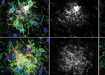
To examine the cellular changes driving this process, the authors utilized a spatial transcriptomics technique called Nova-ST. This advanced method allows scientists to map gene expression across tissue sections while simultaneously visualizing the location of amyloid plaques.
The analysis revealed that microglia located in close proximity to the plaques in Lecanemab-treated mice underwent a distinct transcriptional shift. These cells upregulated genes associated with lysosomes and phagosomes, which are the cellular organelles responsible for ingesting and breaking down waste.
This activation of the digestive machinery was absent in the mice treated with the antibody lacking a functional Fc region. The data suggests that the engagement of the Fc receptor on the microglia by the antibody acts as a switch, turning on a dormant plaque-clearing program.
The researchers further investigated the specific molecular pathways involved using single-cell RNA sequencing. This analysis identified a specific gene network induced by the treatment. One of the most highly upregulated genes was SPP1, which encodes a protein called osteopontin. The expression of this gene was strongly associated with the microglia involved in phagocytosis.
To validate the role of osteopontin, the team performed in vitro experiments using human microglial cultures. They exposed these cells to amyloid plaques on brain tissue sections and added increasing concentrations of the osteopontin protein.
The results showed that higher levels of osteopontin significantly enhanced the ability of the microglia to clear the amyloid deposits. This suggests that the production of osteopontin is a key component of the mechanism by which Lecanemab promotes plaque removal.
A critical concern in Alzheimer’s immunotherapy is the potential for damage to healthy brain tissue, particularly the synapses where neurons communicate. The researchers assessed the density of synaptic markers surrounding the plaques to determine if the increased microglial activity caused collateral damage.
The analysis indicated that Lecanemab treatment did not lead to a reduction in synaptic density. This implies that the induced phagocytosis is specific to the amyloid plaques and preserves the integrity of neuronal connections.
The team also sought to confirm that these findings were not an artifact of the specific mouse model used. They repeated the experiments in mice with intact, fully functional mouse immune systems using a mouse-specific version of the antibody. These groups included eight to nine animals each.
The results mirrored the findings in the humanized model, confirming that the Fc-dependent mechanism is robust and functions in the presence of a complete adaptive immune system.
The study does have certain limitations. The primary mouse model used for the human cell experiments lacked an adaptive immune system, which includes T cells and B cells. While the parallel experiment in immunocompetent mice supports the general conclusions, the complex interactions between microglia and the broader human immune system may influence the therapy’s effects in patients.
Additionally, the animal models used in this research do not typically develop significant vascular pathologies. In human clinical trials, a subset of patients treated with amyloid-clearing antibodies experiences side effects known as amyloid-related imaging abnormalities, which can involve brain swelling or bleeding. The current study could not fully assess how the observed microglial activation might contribute to or interact with these vascular issues.
Future research directions may focus on investigating the diversity of Fc receptors on microglia to understand which specific receptors mediate the beneficial effects versus potential inflammatory side effects.
The identification of the osteopontin pathway also opens new avenues for therapeutic development. It suggests that triggering this specific clearing program, perhaps through small molecules rather than antibodies, could offer an alternative strategy for reducing amyloid burden.
The authors provided a disclosure regarding potential conflicts of interest. Bart De Strooper, the senior author, has served as a consultant for several major pharmaceutical entities, including Eli Lilly, Biogen, Janssen Pharmaceutica, Eisai, and AbbVie. He is also a scientific founder of Augustine Therapeutics and Muna Therapeutics, holding stock in the latter. The remaining authors declared no competing interests.
The study, “The Alzheimer’s therapeutic Lecanemab attenuates Aβ pathology by inducing an amyloid-clearing program in microglia,” was authored by Giulia Albertini, Magdalena Zielonka, Marie-Lynn Cuypers, An Snellinx, Ciana Xu, Suresh Poovathingal, Marta Wojno, Kristofer Davie, Veerle van Lieshout, Katleen Craessaerts, Leen Wolfs, Emanuela Pasciuto, Tom Jaspers, Katrien Horré, Lurgarde Serneels, Mark Fiers, Maarten Dewilde and Bart De Strooper.


