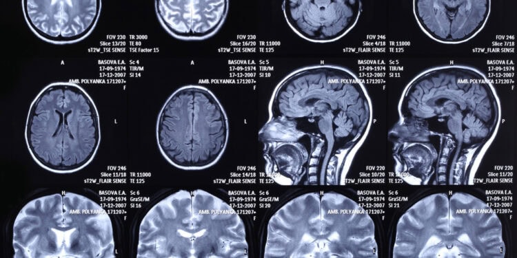
A new brain imaging study provides evidence that high levels of iron in certain parts of the brain may signal an increased risk of developing cognitive problems years before symptoms appear. The study, published in Radiology, found that greater iron accumulation in two brain regions was linked with later development of mild cognitive impairment, a condition that often precedes Alzheimer’s disease. These findings suggest that brain iron could play a role in the early stages of neurodegeneration and might eventually serve as a tool for identifying people at higher risk for memory decline.
Alzheimer’s disease is the most common cause of dementia, gradually impairing memory, thinking, and behavior. It is marked by the buildup of two abnormal proteins in the brain: amyloid-beta and tau. These changes begin silently, sometimes decades before noticeable symptoms. Mild cognitive impairment is often the earliest clinical sign that something is wrong, acting as a transitional phase between normal aging and dementia.
Although researchers have developed methods to detect amyloid and tau, these markers do not fully explain how fast someone will decline. Some people with high amyloid levels never develop dementia, while others deteriorate quickly. This inconsistency has led scientists to explore other contributors to neurodegeneration.
One such factor is iron. Although iron is essential for brain function, too much of it can cause harm. Excess iron may trigger inflammation, damage cells, and worsen the effects of other proteins involved in Alzheimer’s. Earlier studies have shown that iron tends to accumulate with age and that high iron levels may be linked to memory and motor problems. The current study aimed to determine whether measuring brain iron could help predict who might go on to develop cognitive decline, even before any symptoms begin.
“We were motivated by the need for earlier and more practical biomarkers for Alzheimer’s disease. Amyloid and tau are well established Alzheimer’s biomarkers, but are usually detected by costly PET scans, while conventional MRI markers like brain atrophy only appear at late stage,” said senior author Xu Li, an associate professor at Johns Hopkins University School of Medicine and a research scientist in the Kennedy Krieger Institute.
“In addition, therapies targeting amyloid only showed modest effect, suggesting the importance of other co-pathologies. We therefore wanted to test if quantitative susceptibility mapping (QSM) MRI, a MRI method for measuring brain iron, could fill this gap and help identify dementia risks years before cognitive impairment happens.”
Quantitative susceptibility mapping can detect tiny changes in tissue by measuring magnetic properties linked to iron content. The research was part of a larger, long-term project called the BIOCARD study, designed to investigate early signs of Alzheimer’s. The participants were all older adults who showed no signs of memory problems at the beginning.
The scientists focused on ten specific brain regions, including the entorhinal cortex, putamen, hippocampus, and several others involved in memory, language, and motor function. The participants were monitored for up to 7.7 years, during which they received regular cognitive assessments. Some also underwent brain scans to measure amyloid accumulation using positron emission tomography.
By the end of the study, 27 individuals had developed mild cognitive impairment or dementia. The analysis revealed that higher iron levels at the beginning of the study in two regions — the entorhinal cortex and the putamen — were strong predictors of later cognitive decline. Participants with elevated iron in the entorhinal cortex had about twice the risk of developing mild cognitive impairment compared to those with lower levels. Similar patterns were observed for the putamen.
“The key message is that higher brain iron, especially in memory-related regions, was linked to a greater risk of developing memory problems and faster cognitive decline,” Li told PsyPost. “This suggests that brain iron may serve as an early warning sign of Alzheimer’s disease, years before symptoms become obvious.”
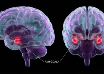
The entorhinal cortex is a region deeply involved in memory and is known to be one of the first areas affected by Alzheimer’s. The putamen, although more commonly associated with movement, also contributes to cognitive processes such as language and flexibility in thinking. Interestingly, these associations between iron levels and future decline were found to be independent of structural brain changes such as shrinkage.
The findings were even more pronounced in individuals who had higher amyloid levels in their brains. Among this group, those with high iron in the entorhinal cortex or putamen showed faster declines in their overall cognitive performance over time. This pattern suggests that brain iron might work in tandem with amyloid buildup to accelerate the disease process.
“We found out that participants with higher brain iron in key memory regions were about twice as likely to develop mild cognitive impairment (MCI), which is a transition stage to dementia,” Li explained. “Such risk could be even higher in participants with known Alzheimer’s pathology such as elevated amyloid burden.”
The study also found that greater iron accumulation in other areas, like the caudate nucleus and hippocampus, was associated with lower cognitive scores over time, particularly in people who were already showing signs of amyloid pathology. In terms of specific mental abilities, higher iron levels in the entorhinal cortex and putamen were linked to declines in language and visuospatial function.
Although the findings are promising, the researchers caution that the results should be interpreted carefully. The study group was relatively small and mostly included highly educated White participants with a family history of Alzheimer’s, which may limit how broadly the findings apply. Additionally, the number of participants who progressed to mild cognitive impairment was modest, especially in the subgroup who underwent both types of brain scans.
Another caveat is that iron accumulation is not unique to Alzheimer’s disease. Elevated brain iron has been seen in other conditions, including Parkinson’s disease and normal aging. This suggests that while brain iron may be a useful marker of neurodegeneration, it may not be specific to Alzheimer’s.
“One potential caveat or misinterpretation I want to point out is that our study does not mean brain iron is the sole cause of Alzheimer’s disease,” Li noted. “It could be a potential early marker of Alzheimer’s related neurodegeneration, but it is likely just one factor among many, and should be considered alongside with other markers such as amyloid and tau. Its exact role, whether causal or secondary, and how it interacts with other Alzheimer’s pathology still need more studies. Anyway, brain iron as measured by QSM MRI could be useful for Alzheimer’s staging, categorizing risk populations and can potentially guide early treatment in the future.”
“Our findings need to be validated in larger and more diverse populations. We want to further investigate how iron interacts with amyloid and tau in Alzheimer’s disease. We are also working on standardizing QSM MRI and hope to make it faster, more reliable, and more widely available in clinical practice.”
“Our study shows that brain iron predicts future MCI risk and memory decline in people who are still cognitively normal,” Li said. “It is among many efforts to develop earlier and better biomarkers for Alzheimer’s disease beyond the well known amyloid and tau pathology. Development of therapies targeting brain iron in Alzheimer’s is still at its very early stage, but hopefully there will be more studies and trials in the future.”
The study, “Susceptibility MRI Helps Predict Mild Cognitive Impairment Onset and Cognitive Decline in Cognitively Unimpaired Older Adults,” was authored by Lin Chen, Anja Soldan, Andreia Faria, Marilyn Albert, Peter C. M. van Zijl, and Xu Li, for the BIOCARD Study Team.


