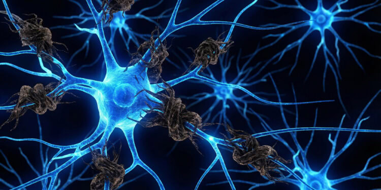

A new study published in the journal Neuropharmacology suggests that chronic alcohol consumption affects the brain differently depending on the specific underlying characteristics of Alzheimer’s disease. The findings indicate that alcohol alters a key brain circuit responsible for decision-making in opposite ways, depending on whether the brain is burdened by amyloid plaques or tau tangles. This provides evidence that the relationship between alcohol use and cognitive decline is highly complex and dependent on a person’s unique biological state.
Alzheimer’s disease is a progressive brain condition traditionally characterized by the buildup of two different proteins. Amyloid-beta forms sticky plaques outside of brain cells, whereas the tau protein creates tangled structures inside the cells. These biological changes disrupt how brain cells communicate, often leading to memory loss and cognitive decline. Aside from these biological markers, lifestyle factors play a role in how the disease progresses.
Alcohol consumption is an established risk factor for dementia, and it is known to alter brain pathways involved in behavioral flexibility and decision-making. Behavioral flexibility is the ability to adjust habits and actions when environmental situations change. A specific neural pathway known as the corticostriatal circuit is largely responsible for this cognitive ability.
The corticostriatal circuit connects the prefrontal cortex to a region called the dorsomedial striatum. The prefrontal cortex handles complex thought and planning, and the striatum manages action and reward processing. This pathway often becomes impaired early in both addiction and Alzheimer’s disease. Postdoctoral researcher Yufei Huang and a team of scientists led by Jun Wang at Texas A&M University wanted to investigate how chronic alcohol use interacts with specific Alzheimer’s-related biological changes.
The researchers aimed to understand if alcohol modifies the corticostriatal circuit differently when amyloid-beta is present compared to when tau tangles are the primary issue. They used two distinct genetic mouse models to represent the different aspects of Alzheimer’s disease. The first model features a genetic modification that causes a rapid accumulation of amyloid-beta plaques in the brain. The second model is genetically engineered to develop tau tangles over time.
Using an intermittent drinking procedure, the researchers provided the mice with a choice between water and a twenty percent alcohol solution. The amyloid-beta mice were exposed to this alcohol routine for 16 weeks starting at two months of age. In a separate experiment, the tau mice began the alcohol routine at six months of age and continued for six months. This timeline matched the natural progression of tau development in that specific genetic model.
After the drinking period concluded, the scientists examined the brain tissue using patch-clamp electrophysiology. This technique involves attaching a microscopic glass pipette to individual brain cells to record their electrical signals. The researchers also used optogenetics, a method that uses light to activate specific, genetically modified nerve endings. To achieve this, they injected a specialized virus into the prefrontal cortex weeks beforehand, which allowed them to stimulate only the specific connection between the prefrontal cortex and the striatum using colored light.
In the amyloid-beta mice, chronic alcohol exposure increased the overall burden of amyloid plaques in the cortex. Alcohol also increased the local electrical activity within the medial prefrontal cortex. The researchers recorded from 35 prefrontal cortex neurons across seven mice and found elevated excitatory signals compared to mice that only drank water.
However, the long-range signals traveling from the prefrontal cortex to the striatum showed a different pattern. When the researchers used light-based stimulation to measure this connection, they observed a significant decrease in communication strength in the alcohol-drinking amyloid mice. The alcohol exposure essentially suppressed the main output of this decision-making circuit in the presence of amyloid plaques.
The tau-based mouse model showed a very different response to the same alcohol exposure. In these mice, alcohol consumption significantly elevated the levels of modified, damaging tau proteins. But local electrical activity in the medial prefrontal cortex remained relatively unchanged compared to tau mice that only drank water.
When examining the long-range connection to the striatum, the scientists found that alcohol actually increased the strength of the communication signals. They recorded from 21 striatum neurons across seven tau mice and observed elevated responses to the light stimulation. This was an unexpected outcome, as the tau protein typically tends to weaken brain connections on its own.
The research team also looked at the brain’s immune cells, which are known as microglia. These cells normally protect the brain by clearing away debris and supporting neuronal health. In the amyloid-beta mice, alcohol exposure caused a significantly higher number of microglia to cluster directly around the amyloid plaques. The tau mice did not show this increased immune cell clustering after alcohol consumption, suggesting a specific immune reaction tied to amyloid buildup.
To test how these immune cells affect brain signals directly, the researchers conducted an additional experiment using wild-type mice without Alzheimer’s traits. They gave three mice an injected drug called PLX5622 for seven days to eliminate most of their microglia. A control group of three mice received a simple saline injection. Recordings from 26 prefrontal cortex neurons across the six total mice showed that removing the immune cells increased the strength of electrical signals. This suggests that immune cells play a direct role in regulating brain activity levels, which may help explain the circuit changes seen in the alcohol-exposed amyloid mice.
Interpreting these findings requires keeping a few limitations in mind. The study relied on animal models, which cannot perfectly replicate the complex progression of human cognitive decline. Humans typically experience a combination of both amyloid and tau pathologies simultaneously, rather than in isolation. Future studies will need to explore how alcohol interacts with a brain state that contains both sticky plaques and tau tangles at the same time.
The genetic backgrounds of the two mouse models were also slightly different, which might naturally influence how heavily alcohol alters these brain pathways. Human environmental exposure is also much more complex than a controlled drinking schedule in a laboratory. Lifestyle factors such as stress, diet, and sleep patterns likely interact with biological markers to shape overall brain health.
The researchers measured electrical activity in brain slices but did not directly test the mice for memory or behavioral deficits. It remains unknown exactly how these specific brain circuit changes translate into real-world confusion or decision-making problems. Future research is expected to incorporate behavioral testing to see if the alcohol-induced brain signal changes directly cause measurable learning impairments in animals.
The study, “Chronic alcohol exposure produces pathology-dependent corticostriatal circuit remodeling in Aβ- and tau-based mouse models of Alzheimer’s disease,” was authored by Yufei Huang, Xueyi Xie, Zhenbo Huang, Himanshu Gangal, Ruifeng Chen, Xuehua Wang, Jianrong Li, and Jun Wang.