
Exposure to elevated levels of sodium fluoride might impair cognitive abilities by altering how brain cells grow and communicate. A new small study of mice and lab-grown tissues shows that excessive amounts of this chemical disrupt the structural proteins necessary for healthy neural connections. The findings were recently published in the journal Brain Research.
Fluoride is an active nonmetal element found widely in the natural world. In small trace amounts, it provides benefits to human health. Dental professionals often recommend low concentrations to harden enamel and prevent tooth decay.
However, excessive exposure over long periods poses systemic health risks. In many regions, surface and groundwater naturally contain elevated levels of the mineral. This occurs when groundwater flows through surrounding rock formations that contain large deposits of fluorite. Communities relying on these untreated water sources can experience chronic fluoride toxicity.
Over time, toxic amounts of the mineral can accumulate in the body. Beyond physical ailments affecting the skeletal framework, high chemical exposures alter the central nervous system. Previous observations in human populations suggest that individuals dwelling in high-fluoride geographic areas often score lower on intelligence tests compared to those living in low-fluoride zones.
The biological mechanisms behind this cognitive decline are largely a mystery to the scientific community, though the topic is drawing increased academic scrutiny. A separate paper published early in 2024 indicated a link between maternal fluoride levels and behavioral issues in toddlers, pointing toward early developmental vulnerabilities. To investigate exactly how the chemical damages neural tissues, lead author Lingli Chen and a team of researchers at the Henan Institute of Science and Technology in China conducted laboratory experiments.
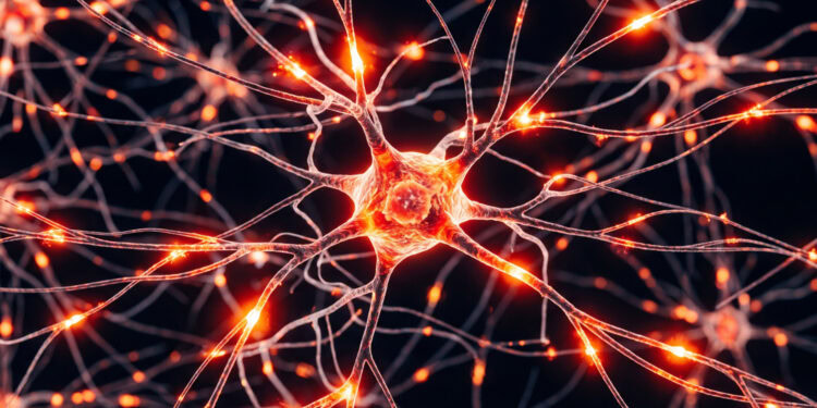
The researchers decided to focus on specific proteins that manage the development and structural stability of brain networks. For the brain to process information and form memories, its nerve cells must communicate effectively. They do this by sending chemical signals across tiny gaps known as synapses.
The receiving and sending ends of these neural connections constantly grow, shrink, and adapt based on an organism’s learning experiences. This adaptable cellular behavior relies on a microscopic internal scaffolding called the cytoskeleton. Several specialized proteins maintain this internal framework and guide the precise formation of new neural branches.
The researchers tracked three prominent structural markers: drebrin, postsynaptic density protein 95, and growth-associated protein 43. Drebrin stabilizes the internal skeleton and helps shape the receiving antennas of neurons. Postsynaptic density protein 95 anchors the receiving end of a synapse, ensuring that signals are caught properly. Growth-associated protein 43 guides the tip of a growing nerve fiber toward its intended target.
To test how fluoride affects these proteins, the research team designed a small study using thirty female mice. After a period of initial acclimation, they divided the animals into three equal experimental groups. One group drank pure deionized water, the second drank water containing fifty milligrams per liter of sodium fluoride, and the third group drank water with one hundred milligrams per liter.
The exposure lasted for five months. Afterward, the team evaluated the learning and memory capabilities of the animals using a behavioral technique known as a step-down test. Researchers temporarily placed the mice on an insulated elevated platform inside a designated testing chamber.
The metallic grid floor surrounding the platform delivered a mild electrical shock. Out of natural instinct, a placed mouse will jump off the elevated stage, but it should quickly learn to stay in place to avoid the uncomfortable floor. The scientists recorded how many times the animals mistakenly jumped down, counting each jump as an error in avoidance memory.
The behavioral results indicated a distinct drop in cognitive performance for the experimental groups. Mice drinking the tainted water experienced weight loss and reductions in overall brain weight. During the testing phase, the animals exposed to the medium dose of fluoride made more errors than those drinking pure water. The highest dose group also made more errors, though the results for that specific comparison were not statistically significant.
Next, the team examined the animal brain tissues using molecular analysis techniques. They measured messenger RNA, which acts as the genetic instructions copied from DNA to build structural proteins. They also measured the final architectural proteins using a laboratory method that identifies specific cellular molecules.
In the animals exposed to the highest levels of fluoride, the genetic instructions for building postsynaptic density protein 95 dropped. The protein levels for drebrin also showed a downward trend after persistent exposure, though the measured changes were not statistically significant.
The blueprints for growth-associated protein 43 also declined as the dose of the chemical increased. Oddly, the physical protein levels for this specific marker actually increased in the low-dose group. The researchers suspect this represents an early bodily compensation mechanism, where the organism attempts to repair initial neural damage by rapidly producing more growth proteins.
To isolate the cellular mechanisms without the interference of a whole animal body, the team conducted a secondary experiment. They utilized a common line of lab-grown cells originally derived from the mouse hippocampus. The hippocampus is a specialized brain region essential for spatial learning and memory consolidation.
The scientists exposed these cultured cells to various concentrations of sodium fluoride, spanning from zero up to fifty millimolar, for twenty-four hours. They observed the samples using scanning laser microscopes. To visualize the microscopic anatomy, they treated the cells with specific fluorescent dyes that bind to the cellular skeleton and light up under the laser equipment.
To ensure their cell counts were precise, the researchers recorded the optical densities of the tissue layers. They found that low doses of half a millimolar had little impact on the cells. However, jumping to two millimolar concentrations prompted a sharp die-off in the artificial environment, halting cell proliferation.
The surviving units exhibited severe physical deformities under the laser microscope. Their branch-like extensions, known as dendrites, began to atrophy. In some samples, the extensions disappeared entirely. The number of connection points between neighboring cells dropped. The internal protein network, particularly a structural component called F-actin, completely lost its dense matrix.
Molecular tests on the cell cultures mirrored the earlier animal results. After a day of exposure, the cells showed a steep decline in the production of drebrin, postsynaptic density protein 95, and growth-associated protein 43. Without these building blocks, the nerve cells simply could not maintain their intended structural shapes.
The results from both the live animals and the isolated cells point toward a disruption in fundamental synaptic structure. By halting the normal production of structural scaffolding materials, excessive fluoride appears to physically degrade the memory networks of the brain. The scientists noted that the specific biological effects depend heavily on the severity of the exposure.
There are a few caveats to consider regarding this particular research approach. The chemical concentrations used in the laboratory experiments were exceptionally high. Natural drinking water rarely approaches the extreme levels applied to the mice and tissues in this setup.
Additionally, while the results demonstrate a strong physiological correlation between toxic exposure and protein degradation, they do not establish an absolute sequential chain of events. The researchers noted that further genetic experiments are necessary to prove causality. In the future, the team plans to use larger samples and environmental concentrations closer to natural human exposures.
The study, “Sodium fluoride exposure induced cognitive impairment via disorders synaptic protein expression and neuronal development in mice brain,” was authored by Lingli Chen, Rui Wang, Penghuan Jia, Qian Jiang, Siyuan An, Zhihong Yin, Dongfang Hu, Hongmei Ning, and Yaming Ge.