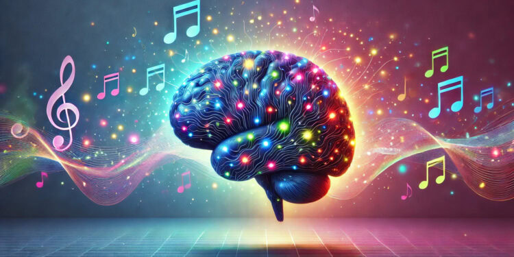

When we listen to music and find it beautiful, our brains engage in a different pattern of activity compared to when we listen to music we don’t find beautiful. A recent study using brain imaging technology discovered that experiencing beauty in music involves increased communication between brain areas associated with reward and visual processing, while listening to music considered less beautiful is linked to more activity in brain regions responsible for basic sensory processing. This suggests that appreciating musical beauty is a complex process that goes beyond just hearing the sounds, involving higher-level brain functions like pleasure and mental imagery.
The research was published in the journal Psychology of Aesthetics Creativity and the Arts.
Humans have a universal tendency to find beauty in things around them and to enjoy this experience. This ability to appreciate beauty is thought to be deeply rooted in our biology and has likely played a role in human evolution. Music is one of the most common sources of beauty in our lives, with people around the world spending a significant amount of time listening to it.
While we know that judging music as beautiful or not is a common and seemingly natural human behavior, the brain processes behind this aesthetic judgment are not fully understood. Previous research has identified some key brain regions that become active when we make aesthetic judgments, particularly a region called the orbitofrontal cortex, which is involved in evaluating rewards.
However, these studies often focused on activity in specific areas and did not fully explore how different brain regions communicate with each other over time during the experience of musical beauty. Researchers wanted to take a closer look at these dynamic brain connections to understand what happens in the brain when we listen to music and decide whether it is beautiful or not. They used a new method that allows them to track changes in brain connectivity as they happen over time, hoping to reveal more about the neural mechanisms behind our aesthetic experiences of music.
To explore this question, researchers recruited 36 healthy adults with varying levels of musical background. During the study, participants lay in a brain scanner that uses functional magnetic resonance imaging, or fMRI. This technology measures brain activity by detecting changes in blood flow. While in the scanner, participants listened to a piece of music called “Adios Nonino” by Astor Piazzolla. This piece was chosen because it contains a lot of musical variety, including contrasting sections, and previous studies had shown that people tend to have consistent opinions about which parts are beautiful.
In a separate session, with a different group of people, the researchers had participants continuously rate the perceived beauty of the same Piazzolla music piece. They used a motion sensor, similar to a video game controller, to allow participants to indicate in real-time whether they found the music beautiful or not. By analyzing these ratings, the researchers were able to identify specific musical passages that were consistently rated as “beautiful” and other passages consistently rated as “non-beautiful.”
After identifying these beautiful and non-beautiful musical segments based on the ratings, the researchers went back to the brain scan data from the participants who had listened to the music in the scanner. They used a sophisticated analysis technique called Leading Eigenvector Dynamics Analysis to examine how different brain regions communicated with each other while participants listened to the music. This method is designed to capture the changing patterns of brain connectivity as they occur over time, rather than just looking at average connectivity over the entire listening period.
The analysis identified recurring patterns of brain communication, which the researchers called “functional connectivity states.” They then compared how often these different brain connectivity states occurred when participants were listening to the musical passages rated as beautiful versus when they were listening to the passages rated as non-beautiful. They also looked at how frequently the brain switched between these different connectivity states in both conditions. This allowed them to see if there were specific brain network patterns and transitions associated with experiencing musical beauty.
The researchers identified 12 distinct, recurring patterns of brain connectivity that occurred while participants listened to the music. When they compared the occurrence of these states during beautiful versus non-beautiful music listening, they found that three states showed significant differences. One brain connectivity state, which involved visual areas of the brain located at the back of the head, was more frequently observed when participants listened to music rated as beautiful. These visual areas are typically associated with processing what we see, but they can also be involved in mental imagery and visualization.
In contrast, two other brain connectivity states were more frequent when participants listened to music rated as non-beautiful. One of these states primarily involved auditory brain regions, specifically areas responsible for basic sound processing, located in the temporal lobes near the ears. The other state that was more common during non-beautiful music listening was more complex, involving not only auditory areas but also brain regions related to movement control, sensory processing, and emotion processing, such as the amygdala, known for its role in processing emotions like fear.
Furthermore, the researchers examined how frequently the brain transitioned between these different connectivity states. They discovered that during listening to beautiful music, there were more frequent transitions involving a network that included the orbitofrontal cortex (the reward evaluation area), visual areas, and regions related to reward processing. This suggests that when we experience musical beauty, there is a dynamic interplay and rapid communication between brain regions involved in reward, visual imagery, and overall evaluation.
Conversely, during listening to non-beautiful music, there were more frequent transitions involving brain states related to auditory processing and regions associated with emotional responses, such as the amygdala and insula. This might indicate that when we listen to music we don’t find beautiful, our brains are more focused on processing the auditory information itself and dealing with potentially negative emotional responses.
While these findings offer valuable insights into the brain networks involved in aesthetic musical experiences, the researchers acknowledge some limitations. This study was exploratory, using a relatively new method for analyzing brain connectivity, and the statistical differences observed were not very strong after correcting for multiple comparisons. This means that while the results are suggestive and interesting, they should be interpreted with some caution and need to be confirmed in future studies.
Future research should investigate the robustness of these findings using larger groups of participants, different musical pieces, and further explore the capabilities of this dynamic brain connectivity analysis technique. Moving forward, researchers aim to further unravel the complex and dynamic interplay of brain networks that contribute to our rich and varied aesthetic experiences with music, and how these networks shift and communicate as we listen and judge music’s beauty.
The study, “Beauty Is in the Brain Networks of the Beholder: An Exploratory Functional Magnetic Resonance Imaging Study,” was authored by Ruijiao Dai, Petri Toiviainen, Peter Vuust, Thomas Jacobsen, and Elvira Brattico.


