![[Adobe Stock]](https://sp-ao.shortpixel.ai/client/to_webp,q_glossy,ret_img,w_750,h_375/https://www.psypost.org/wp-content/uploads/2025/04/man-touching-his-hair-750x375.jpg)
A study of healthy adult men found that elevated testosterone levels were detectable in hair seven weeks after the transdermal administration of a single dose of testosterone. Similarly, elevated cortisol levels—a hormone associated with stress—were observed in hair samples seven weeks after participants underwent an acute laboratory stressor. The study was published in Psychoneuroendocrinology.
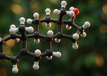
Steroid hormones are a class of hormones derived from cholesterol that play essential roles in regulating metabolism, immune function, and sexual and reproductive development. Examples include estrogen and progesterone, primarily in females; testosterone, primarily in males; and cortisol and aldosterone, which are involved in stress responses and blood pressure regulation, respectively.
These hormones are primarily produced by the adrenal glands and gonads (ovaries in females and testes in males). They exert their effects by entering cells and binding to specific receptors in the cell nucleus, where they influence gene expression and cell activity. Because of their broad physiological influence, imbalances in steroid hormone levels can lead to a range of health issues, including metabolic disorders, cardiovascular disease, and reproductive dysfunction.
Steroid hormones can accumulate in hair, which has led to the growing use of hair analysis in research as a way to assess long-term hormonal exposure. Hormone concentrations in hair can reflect ongoing biological conditions, including chronic stress, the use of hormonal contraceptives, and even the consumption of illicit substances.
Study lead author Hana H. Kutlikova and her colleagues set out to determine whether a one-time testosterone dose could be detected in hair weeks later and whether an acute laboratory-induced stressor would elevate hair cortisol levels. Cortisol is a hormone released by the adrenal glands in response to stress.
The study included 88 healthy adult men between the ages of 18 and 40, with an average age of 24. About 47% of the participants had straight hair and 52% had wavy hair.
At their initial laboratory visit, participants provided cheek cell samples for genetic testing and a hair sample approximately 2–3 mm in circumference taken from the upper back of the scalp (the posterior vertex). Hormone levels in hair were later analyzed using a method called liquid chromatography-tandem mass spectrometry. Saliva samples were also collected.
Participants were randomly assigned to receive either 150 mg of testosterone or a placebo in the form of a topical cream. Neither the participants nor the experimenters knew which cream was administered. Participants applied the cream to their upper arms and shoulders under supervision and then waited for two hours while completing personality questionnaires and engaging in leisure activities.
Following this, participants were randomly assigned to one of three experimental conditions:
- Cold-pressor test (somatic stressor): Participants immersed their hand and wrist into ice-cold water and were asked to keep it there as long as possible (up to five minutes).
- Socially evaluated cold-pressor test (social-somatic stressor): The same procedure was used, but participants were also told they would be video recorded for facial expression analysis. A female experimenter sat facing them and maintained constant eye contact, instructing them to look into the camera throughout.
- Control condition: Participants immersed their hand in lukewarm water and could withdraw it at any time.
Seven weeks after the initial visit, participants returned to the lab and provided another hair sample from a location adjacent to the original site.
The results showed that participants who underwent the cold-pressor test had significantly elevated cortisol levels in their hair seven weeks later. Similarly, those who received testosterone cream had elevated hair testosterone levels compared to those who received the placebo.
“Our study provides new insights into the sensitivity of hair analysis for detecting hormonal changes following single-dose hormone administration and experimentally induced short-term stress events. The exploratory findings emphasize the importance of individual contextual factors in influencing hair hormone concentrations and lay the groundwork for further investigation into the dynamics of cumulative hair hormone measurements,” the study authors concluded.
The study sheds light on the accumulation of hormones in hair. However, it should be noted that the study was conducted exclusively on men. Results on women might not be identical. Additionally, the researchers did not control for certain contextual variables that could influence hormone concentrations in hair, such as physical activity, environmental exposures, or hair treatment practices.
The paper, “The effects of single testosterone administration and stress induction on steroid hormone levels in hair,” was authored by Hana H. Kutlikova, Christoph Eisenegger, Aniko Krumbholz, Igor Rieˇcanský, Claus Lamm, and Boris B. Quednow.


