![Illustration of brain regions studied in mental illness: ACC, amygdala, hippocampus, prefrontal cortex. [NIH]](https://sp-ao.shortpixel.ai/client/to_webp,q_glossy,ret_img,w_750,h_375/https://www.psypost.org/wp-content/uploads/2015/08/Hippocampus-prefrontal-cortex-amygdala-anterior-cingulate-cortex-by-NIMH-750x375.jpg)
New insights into the brain could pave the way for targeted treatments for anxiety, depression, and other mental health conditions. In a groundbreaking study, scientists at the University of California, Davis, have identified distinct groups of cells within the amygdala, a brain region central to emotional processing. By mapping the cellular and genetic structure of the amygdala in humans and primates, researchers hope to unlock new, cell-specific approaches to mental health treatment. The findings have been published in the American Journal of Psychiatry.
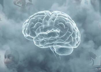
The amygdala is a small, almond-shaped structure located deep within the brain’s temporal lobe, playing a central role in processing emotions, particularly fear and stress responses. This part of the brain helps regulate emotional reactions to different stimuli, sending signals that influence how we react to potentially dangerous or threatening situations.
Given its role in emotional processing, the amygdala has been a focus in studies on various mental health conditions, from anxiety and depression to autism spectrum disorders. Researchers have found that dysregulation in amygdala functioning may contribute to these disorders, highlighting its importance in understanding emotional and behavioral health.
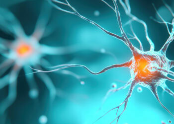
Despite its significance, treating amygdala-related disorders has been challenging because this brain region is far more complex than initially understood. The amygdala contains many different cell types, each with distinct functions and gene expression patterns that influence how it responds to various stimuli.
Most prior research has either focused on the amygdala as a single functional unit or relied on animal models to study its cells, often overlooking the cellular differences within specific areas. As a result, treatments targeting the amygdala’s activity have had limited success, potentially because they did not address the specific cell types involved in emotional dysregulation.
This study was motivated by a need to address this complexity and refine our understanding of the amygdala at the cellular level, especially in humans. The researchers aimed to identify and categorize the different cell types within the amygdala, using gene expression patterns to understand how each cell cluster might contribute to emotional and psychological functions.
“Decades of research has implicated the amygdala in many forms of psychopathology and neurodevelopmental disorders. However, large-scale studies have found these associations to be relatively weak, and effective amygdala-focused treatments have remained elusive,” said study author Andrew S. Fox, an associate professor at UC Davis and core scientist in neuroscience & behavior at the California National Primate Research Center.
“At the same time, basic science research in rodents has revealed many different cell types within the amygdala, which can have distinct or even opposing roles in behavior. This cell-type variability can help explain why effective treatments that target the amygdala have been so hard to find. In short, treatments should not target the amygdala; they should target specific cells in the amygdala. In our paper, we explored the cell-type composition of the amygdala in humans and nonhuman primates to help bridge this gap in our understanding.”
A primary tool used in the research was single-nucleus RNA sequencing, a method that enables scientists to examine the activity of thousands of genes within individual cells. This method was selected because it allows a close look at cell diversity.
The study’s sample included six postmortem subjects—three humans and three rhesus monkeys. Rhesus monkeys were chosen because their amygdala structures and gene expressions closely resemble those of humans, making them a valuable model for understanding human brain function. Brain tissue samples were taken from two specific areas within the amygdala: the central amygdala and the ventral lateral amygdala.
These two regions were selected because they have distinct functions and structural compositions, with the central amygdala being primarily composed of inhibitory neurons involved in processing fear and stress responses, and the ventral lateral amygdala containing mainly excitatory neurons that contribute to emotional learning and memory. Using RNA sequencing, researchers could measure which genes were active in these regions and map out the cellular makeup of each part of the amygdala.
After sequencing, the data were processed to identify clusters of cells with similar gene expression profiles. This analysis revealed several distinct cell clusters within each region of the amygdala, highlighting the complexity and diversity of its cellular structure. Each cluster represented a group of cells with shared gene expressions, which are likely linked to specific functions within the amygdala.
In the central amygdala, for instance, the researchers found a higher concentration of inhibitory neurons, which are cells that suppress brain activity, suggesting their role in regulating emotional responses and keeping fear and stress reactions in check. In contrast, the ventral lateral amygdala contained more excitatory neurons, which are cells that stimulate brain activity and are thought to contribute to learning and memory, particularly emotional memories.
“Brain regions like the amygdala are complex,” Fox told PsyPost. “They contain multiple subregions and many distinct cell types. Scientists are beginning to make progress in understanding what cell types are most relevant to mental illness. This understanding has the potential to guide the development of new behavioral and pharmacological treatments that could provide much-needed relief to people who are suffering.”
One notable finding was the identification of a specific cell cluster in the ventral lateral amygdala that showed gene markers commonly associated with autism spectrum disorder. This cell type, unique to the ventral lateral amygdala, suggests that certain cells within this brain region may be involved in neurodevelopmental disorders.
In the central amygdala, another cell cluster was found to express high levels of genes linked to anxiety and depression. This cluster consisted of intercalated cells, which are small groups of neurons that act as gatekeepers for signals within the amygdala. In both humans and macaques, these intercalated cells expressed the FOXP2 gene and other markers like TSHZ1 and were located along the boundaries of the central amygdala. In previous research with rodents, FOXP2-expressing intercalated cells acted as “gatekeepers,” controlling signals entering and exiting the amygdala.
This discovery is significant because it suggests that specific cell types within the amygdala, rather than the whole structure, may provide a biological basis for anxiety and depression. The researchers propose that these intercalated cells could be a promising avenue for treatment development, with their expression of FOXP2 and additional features—such as the expression of Neuropeptide FF Receptor 2 (NPFFR2), a receptor that could be targeted pharmacologically—making them a potential target for precision therapies.
“We were surprised to identify intercalated cells as being particularly important for anxiety,” Fox explained. “Rodent work has identified this small population of intercalated cells to be important for threat processing, but we know very little about these cells in humans and other primates. By focusing on these cell types we were further surprised to find relatively high levels of expression of an understudied receptor for Neuropeptide FF, which could be explored in relation to treating psychopathology.”
While this study is promising, it has limitations. The sample size is small, and the study was limited to male subjects, which may not fully capture sex-based differences in the amygdala’s structure and function. Additionally, while rhesus monkeys are genetically close to humans, differences between species could influence the study’s applicability. Future research could explore these variations in larger and more diverse samples, possibly expanding to female subjects and exploring more brain regions.
“This research is on the cutting edge, and we should be wary of over-interpreting our findings before replicating them,” Fox cautioned. “In addition, there is a tendency for people to focus on finding a single cure for complex disorders—I do not think such a cure exists. Rather, we must appreciate the complexity of the brain and put our efforts toward identifying potential treatments that can help even a small fraction of those suffering. If scientists work together, these treatments can add up to have a huge impact on the lives of people who are suffering across the world.”
Nevertheless, the research opens new pathways for studying brain disorders by providing a clearer map of the amygdala’s cellular structure in both humans and primates. The work sets the foundation for future research to examine the amygdala’s cell types and functions in greater detail.
“The goal of this research is to lay the foundation for a refined understanding of the amygdala that can help bridge the gap between our cellular and molecular understanding of the rodent brain and the human condition,” Fox told PsyPost. “This work reflects an effort to increase our understanding of what the amygdala actually is. Our hope is that this understanding will guide further research in rodents and humans that is aimed at decreasing suffering.”
“We believe that the translational approach outlined in this manuscript represents an important first step in linking cellular and molecular knowledge gleaned from rodent studies to human psychopathology. A major challenge in the coming years will be to incorporate our increasing knowledge of cell types into modern theories of psychiatric and neurodevelopmental disorders.”
The study, “Translational Insights From Cell Type Variation Across Amygdala Subnuclei in Rhesus Monkeys and Humans,” was authored by Shawn Kamboj, Erin L. Carlson, Bradley P. Ander, Kari L. Hanson, Karl D. Murray, Julie L. Fudge, Melissa D. Bauman, Cynthia M. Schumann, and Andrew S. Fox.


