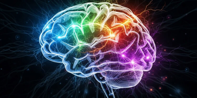
Sleep is often said to help the brain “lock in” what we learn while awake, but the underlying biology remains a topic of debate. A new study published in The Journal of Neuroscience provides evidence that sleep spindles — brief bursts of brain activity that occur during light non-REM sleep — may help reinforce motor memories by targeting the specific brain areas used during learning.
The findings suggest that sleep spindles are not random but instead follow a targeted pattern that supports learning. Rather than occurring uniformly across the brain, spindles appear to concentrate in cortical areas activated during a task, potentially making them a better indicator of memory consolidation than general spindle activity.
The study was led by Martin Sjøgård and Dimitrios Mylonas from Massachusetts General Hospital and Harvard Medical School, alongside an interdisciplinary team of scientists. Their research focused on sleep spindles, which are believed to play a central role in stabilizing and integrating new memories, especially those involving skills and procedures.
Previous studies have shown that these bursts can occur throughout the cortex, but they often appear more prominently in certain regions depending on what was learned while awake. The team wanted to examine whether these bursts of activity are shaped by prior learning, and whether their location in the brain can predict how much a person’s performance improves after sleep.
To do this, they used two complementary brain imaging techniques—electroencephalography and magnetoencephalography—which, when combined with magnetic resonance imaging, allowed them to localize spindle activity across the brain’s surface with much higher spatial precision than conventional methods. This approach provided a more detailed view of how the sleeping brain revisits and potentially strengthens memories of recent experiences.
For their study, the researchers recruited 25 healthy adults and recorded their brain activity during three separate nap visits, spaced at least a week apart. One session was used for adaptation, another served as a baseline, and the final one followed motor learning. During the motor learning visit, participants performed a finger-tapping task known as the motor sequence task. In this task, participants repeatedly typed a five-digit sequence using their left hand. They completed twelve 30-second trials, with rest breaks between each trial. Their performance was measured by the number of correct sequences typed per trial, which captured both speed and accuracy.
Participants then took a 90-minute nap inside the MEG scanner. Brain activity during non-REM Stage 2 sleep was analyzed for spindle events. A few hours later, participants returned to the scanner and were tested again on the same finger-tapping task to measure how much they had improved.
To pinpoint which brain areas were active during learning, the researchers first examined frequency changes in power from rest to task performance. They identified a network of 102 cortical regions that showed significant suppression of activity during task performance, which included primary motor and somatosensory areas, premotor regions, and supplementary motor areas.
The main finding was that sleep spindle density increased in these task-engaged regions after learning compared to baseline sleep. Specifically, about 18 percent of the identified task-related regions showed a learning-induced increase in spindle activity, compared to just under 3 percent of regions outside of this network. These regions included bilateral hand areas of the primary motor cortex, motor planning regions, and supplementary motor areas.
Importantly, these increases were not arbitrary. Spindle density changes predicted how much participants improved on the task after the nap. But this relationship was region-specific. Performance gains following sleep were associated with increased spindle activity in areas involved in motor planning, such as the supplementary motor area and contralateral premotor cortex.
In contrast, improvements during training itself were linked to spindle activity in motor execution areas like the primary motor cortex and somatosensory regions. The two sets of regions did not overlap, suggesting that initial learning and sleep-dependent improvement rely on different neural processes.
This distinction adds weight to the idea that learning during wakefulness and improvement after sleep reflect separate components of memory formation. Learning may encode the experience, while sleep consolidates and refines it. The study authors suggest that spindles in execution-related areas might stabilize the memory trace, while spindles in planning areas may support the refinement and automation of the skill during sleep.
The researchers also analyzed whether participants who naturally had higher spindle density during the nap were simply better learners. They found that spindle density during the learning-related nap predicted better performance, but spindle density during the baseline nap did not. This suggests that it was the learning experience itself that triggered the increase in spindle activity, rather than individual differences in baseline spindle production.
Interestingly, the study also found increases in spindle activity in regions not directly related to hand movement. For example, areas of the motor cortex involved in mouth and face control also showed heightened spindle density after learning. This may relate to participants’ reported use of covert verbal rehearsal during the task, as many silently repeated the number sequence to guide their movements. Previous work has suggested that these regions may contribute to motor planning and verbal rehearsal, and the current results support the idea that their activation may influence memory consolidation as well.
The authors note that these findings provide support for a growing view that spindles are not a generic sleep phenomenon but a targeted one. Their focal expression may allow the brain to selectively strengthen specific neural circuits, depending on what was learned. This might help explain why deficits in spindle production are observed in conditions like schizophrenia and autism, which are marked by difficulties with learning and memory.
The study relied on daytime naps, during which lighter sleep stages such as non-REM Stage 2 predominate. The team chose to focus on this stage because spindles are more frequent there and have been more strongly linked to motor memory improvements. Only a few participants experienced deep slow-wave sleep during the nap, so the role of this stage remains unclear and would need to be explored in future overnight studies.
Another limitation is that the researchers focused exclusively on a motor task, so the generalizability to declarative or emotional memory tasks remains to be tested. Still, the authors suggest that similar principles may apply. If other types of learning also recruit spindles in specific cortical regions, then tracking these changes could provide a sensitive marker of how well the brain is consolidating new information.
Looking ahead, the research team aims to apply this framework to understand how learning and memory processes are affected in neurodevelopmental disorders. Because spindles are measurable and can be enhanced through noninvasive stimulation techniques, this work may eventually inform new treatments to boost learning in clinical populations.
The study, “Increased Sleep Spindles in Regions Engaged during Motor Learning Predict Memory Consolidation,” was authored by Martin Sjøgård, Dimitrios Mylonas, Bryan Baxter, Zhaoyue Shi, Sheraz Khan, Charmaine Demanuele, Lin Zhu, Catherine Tocci, Robert Stickgold, Matti S. Hämäläinen, and Dara S. Manoach.


