![[Adobe Stock]](https://sp-ao.shortpixel.ai/client/to_webp,q_glossy,ret_img,w_750,h_375/https://www.psypost.org/wp-content/uploads/2025/01/neurons-1-750x375.jpg)
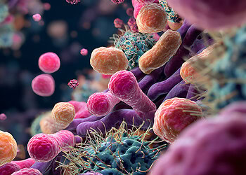
A new study published in eLife has found that a remarkably rare type of neuron acts as a master conductor for the brain’s electrical activity and blood flow. Researchers at The Pennsylvania State University found that removing these specific cells, known as type-I nNOS neurons, from one side of a mouse’s brain led to significant disruptions in both local brain function and the synchronized communication between the two cerebral hemispheres.
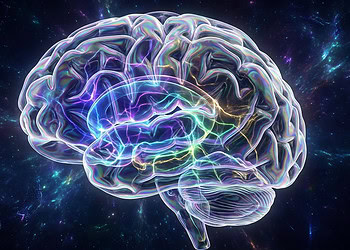
The work suggests this sparse population of cells plays an outsized role in orchestrating brain-wide dynamics, and their loss could contribute to the cognitive decline associated with aging and neurodegenerative disease.
The brain is a profoundly complex and energy-demanding organ. To function properly, its billions of neurons must constantly communicate through electrical signals, a process that requires a steady and precisely regulated supply of oxygen and nutrients from the bloodstream. This tight relationship between neural activity and blood flow is known as neurovascular coupling. When a group of neurons becomes active, nearby blood vessels dilate to increase blood supply to that region, a fundamental process that forms the basis of modern brain imaging techniques like functional magnetic resonance imaging.
One of the key chemical messengers involved in dilating these blood vessels is nitric oxide. This small molecule is produced by an enzyme called neuronal nitric oxide synthase, or nNOS, which is found in certain inhibitory brain cells called interneurons.
Researchers have identified two main categories of these cells, but a team of engineers and neuroscientists led by Patrick J. Drew at Penn State focused on a particularly enigmatic subgroup called type-I nNOS neurons. These cells are exceedingly scarce, making up less than one percent of all neurons in the cortex, yet they possess long, branching connections that extend across wide swaths of the brain. The researchers wanted to understand what specific functions these rare but widely connected cells perform.
A defining feature of type-I nNOS neurons is that they are the only cells in the cortex that have a specific surface receptor, called TACR1. This receptor acts like a lock that can only be opened by a particular key, a molecule called substance P. The research team used this unique feature to design a highly specific method for studying the neurons’ function.
They engineered a “molecular weapon” by attaching a potent toxin, saporin, to a synthetic version of substance P. When this compound was injected into a small region of the mouse brain, only the type-I nNOS neurons with the TACR1 receptor would absorb it, leading to their selective elimination while leaving other neighboring cells unharmed.
The team injected this substance into the somatosensory cortex of mice, the brain region responsible for processing touch, particularly from the whiskers. A separate group of control mice received an injection with a harmless, scrambled version of the substance P-toxin conjugate.
After allowing several weeks for recovery, the scientists used an array of advanced techniques to observe brain function in the awake, behaving animals. Widefield optical imaging allowed them to measure changes in blood volume across the brain’s surface, while two-photon microscopy provided a magnified view of individual arteries dilating and constricting. At the same time, implanted electrodes recorded the collective electrical activity of neurons, known as the local field potential.
When the researchers puffed air on the mice’s whiskers to simulate a touch sensation, they observed notable changes in the brain’s response. In mice lacking the type-I nNOS neurons, the sustained increase in blood flow during a prolonged stimulus was significantly reduced. After a brief stimulus, the typical small dip in blood volume that follows the initial surge was completely absent. These results indicated that while these neurons are not solely responsible for initiating blood flow responses, they play a substantial part in shaping and sustaining them over time.
The elimination of these neurons also had a profound effect on the brain’s intrinsic electrical rhythms. The researchers found a marked reduction in the power of slow brain waves in the delta frequency band, between 1 and 4 Hertz. These slow waves are prominent during deep sleep and are thought to be important for memory consolidation and the brain’s waste clearance system. The reduction in delta wave activity was apparent across all states, whether the animals were alert, quietly resting, or asleep.
Beyond these local effects, the study revealed a breakdown in brain-wide coordination. Normally, the corresponding regions of the brain’s left and right hemispheres show highly synchronized activity. However, after removing the type-I nNOS neurons from just one hemisphere, this synchrony diminished.
The moment-to-moment fluctuations in blood volume and the patterns of high-frequency neural activity became less correlated between the two sides of the brain. This suggests that these rare neurons act as key nodes in a network that helps bind the two hemispheres together, ensuring they operate in a coordinated fashion.
Another key finding related to the spontaneous, rhythmic pulsing of the brain’s arteries, a phenomenon known as vasomotion. These oscillations, which occur even in the absence of any specific task or stimulation, are believed to help circulate cerebrospinal fluid and clear metabolic waste products from the brain. In mice without type-I nNOS neurons, the amplitude of these resting-state blood volume oscillations was significantly dampened. The arteries still pulsed, but their rhythmic dilations and constrictions were much weaker.
The study includes some important considerations. The experiments were conducted weeks after the neurons were removed, which may have allowed the brain to partially reorganize and compensate for their absence. Additionally, the relationship between neural signals and vascular responses is not always linear.
It is possible that multiple redundant pathways for vasodilation exist, such that removing one component does not cause a complete failure of the system, especially during strong stimulation when the response might already be near its maximum. This could help explain why some previous studies that artificially activated these neurons saw very large vascular effects, while this study saw more nuanced changes upon their removal.
Future research will likely explore the long-term consequences of losing these specific neurons. Because type-I nNOS neurons are known to be particularly vulnerable to cellular stress, their gradual loss over a lifetime could be a contributing factor to age-related cognitive decline. By connecting this select population of cells to fundamental processes like delta waves, interhemispheric communication, and vasomotion, this work opens up new avenues for understanding how the brain maintains its health and how these systems can fail in disease.
The study, “Type-I nNOS neurons orchestrate cortical neural activity and vasomotion,” was authored by Kevin Turner, Dakota Brockway, Md Shakhawat Hossain, Keith Griffith, Denver Greenawalt, Qingguang Zhang, Kyle Gheres, Nicole Crowley, and Patrick J Drew.


