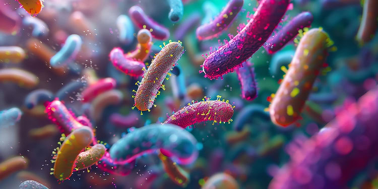
A new study published in Nature Structural and Molecular Biology offers groundbreaking evidence that gut bacteria can influence how proteins in the brain are chemically modified by sugars, a process known as glycosylation. Using a new method called DQGlyco, the researchers were able to analyze brain glycosylation with unprecedented resolution, revealing that different microbial populations in the gut can alter these patterns in ways that may affect brain function.
Protein glycosylation is a biological process in which sugar molecules are added to proteins, helping regulate how proteins function, interact, and communicate within and between cells. While this process plays important roles in many systems of the body, its involvement in the brain has been difficult to study due to the complexity and diversity of sugar modifications. Past studies have also been limited by technical constraints in identifying and quantifying the many forms of glycosylation that can occur on a single protein.
To overcome these challenges, researchers developed a method called deep quantitative glycoprofiling, or DQGlyco. This new technique allows scientists to measure protein glycosylation at a scale and resolution not previously possible. The team then used DQGlyco to examine how glycosylation patterns in the mouse brain are affected by different gut microbiomes.
The study began by validating the DQGlyco method in both human cells and mouse brain tissue. This method combines improved sample preparation, high-sensitivity detection, and precise labeling techniques to isolate and analyze glycopeptides—protein fragments with attached sugar chains. The researchers applied their method to brain tissue from mice, identifying over 177,000 unique glycopeptides. This is more than 25 times the number identified in previous studies, offering an unprecedented view of the brain glycoproteome.
With this enhanced dataset, the researchers discovered widespread “microheterogeneity”—meaning that individual sites on a protein often carried many different sugar forms, known as glycoforms. On average, they found about 17 glycoforms per site, and in some cases, more than 600 different glycoforms at a single location. This variation was not random. Sites with greater microheterogeneity were more likely to be located in structured and accessible parts of the protein, suggesting that structural features help determine how extensively proteins are glycosylated.
The study also revealed that certain types of glycosylation—such as those involving specific sugar types like fucose or sialic acid—were more common on particular protein domains, such as those involved in immune signaling or cell adhesion. These findings suggest that the chemical diversity of sugar modifications may be functionally relevant, influencing how proteins interact and perform their roles.
To explore the influence of the gut microbiome on brain glycosylation, the team conducted experiments using germ-free mice. These mice were either kept sterile or colonized with different bacterial populations, including a single common gut species or a defined community of eight bacteria. Two weeks later, the researchers analyzed the mice’s brain tissue.
The results showed substantial differences in glycosylation patterns between the groups. Over 2,500 glycopeptides in the brain were affected by the presence or absence of gut microbes. These changes were especially prominent on proteins known to be involved in neurotransmission, such as glutamate receptors and ion channels. Many of the altered glycoforms were located at sites with high microheterogeneity, indicating that microbial signals might be influencing the most dynamically glycosylated regions of brain proteins.
Interestingly, only a small number of changes in protein abundance were observed, suggesting that the differences were not due to changes in how much protein was produced, but rather in how those proteins were modified after being made. Additional experiments showed that glycosylation changes were also associated with differences in protein solubility and thermal stability, two indicators of changes in protein function or localization.
The study also found that certain glycoforms were more likely to be surface-exposed on cells, using enzymes to selectively cleave sugars or proteins from intact cells. Surface-exposed glycoforms tended to be more complex and processed, whereas high-mannose forms—often considered immature—were less accessible. This helped distinguish between proteins that were still being processed inside cells versus those functioning at the cell surface.
By combining deep profiling with structural predictions from AlphaFold, the researchers created a model that could predict whether a given glycosylation site was likely to be highly variable or not. This model, based on structural exposure and folding, successfully distinguished between high and low microheterogeneity sites and suggested that glycosylation patterns are influenced by the 3D structure of proteins.
While the findings open new directions for understanding the gut-brain axis, the study has several limitations. The experiments were conducted in mice, so it is not yet clear how directly the results apply to humans. The study also measured glycosylation patterns at a specific time point, meaning that the dynamic nature of these changes over longer periods remains unknown. Additionally, although changes in glycosylation were linked to structural and biophysical changes in proteins, the functional consequences for behavior or brain activity were not directly tested.
Future research may explore how these glycosylation changes influence neural development, cognition, or disease risk. The authors also suggest that their DQGlyco method could be applied to human tissue or disease models to identify glycosylation signatures linked to neurological or psychiatric disorders.
The study, “Uncovering protein glycosylation dynamics and heterogeneity using deep quantitative glycoprofiling (DQGlyco),” was authored by Clément M. Potel, Mira Lea Burtscher, Martin Garrido-Rodriguez, Amber Brauer-Nikonow, Isabelle Becher, Cecile Le Sueur, Athanasios Typas, Michael Zimmermann, and Mikhail M. Savitski.