
Recent analysis of human brain tissue suggests that a small and often overlooked region deep within the brain may play a central role in bipolar disorder. Researchers found that neurons in the paraventricular thalamic nucleus are depleted and genetically altered in people with the condition. These results point toward potential new targets for diagnosis and treatment. The findings were published in the journal Nature Communications.
Bipolar disorder is a mental health condition characterized by extreme shifts in mood and energy levels. It affects approximately one percent of the global population and can severely disrupt daily life. While medications such as lithium and antipsychotics exist, they do not work for every patient. These drugs also frequently carry difficult side effects that cause patients to stop taking them. To develop better therapies, medical experts need a precise map of what goes wrong in the brain.
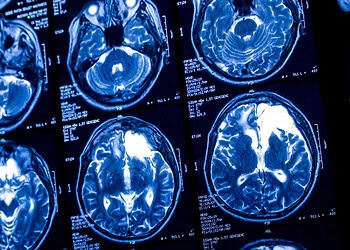
Past research has largely focused on the outer layer of the brain known as the cortex. This area is responsible for higher-level thinking and processing. However, brain scans using magnetic resonance imaging have hinted that deeper structures also shrink in size during the course of the illness. One such structure is the thalamus. This central hub acts as a relay station for sensory information and emotional regulation.
Within the thalamus lies a specific cluster of cells called the paraventricular thalamic nucleus. This area is rich in chemical messengers and has connections to parts of the brain involved in emotion. Despite these clues, the molecular details of this region remained largely unmapped in humans. A team led by Masaki Nishioka and Tadafumi Kato from Juntendo University Graduate School of Medicine in Tokyo launched an investigation to bridge this gap. They collaborated with researchers including Mie Sakashita-Kubota to analyze postmortem brain tissue.
The researchers aimed to determine if the genetic activity in this deep brain region differed from healthy brains. They examined brain samples from 21 individuals who had been diagnosed with bipolar disorder and 20 individuals without psychiatric conditions. They looked at two specific areas: the frontal cortex and the paraventricular thalamic nucleus. To do this, they used a technique called single-nucleus RNA sequencing.
This technology allows researchers to catalog the genetic instructions being used by individual cells. By analyzing thousands of nuclei, the team could identify different cell types and see which genes were active or inactive. This provided a high-resolution view of the cellular landscape. They compared the data from the thalamus against the data from the cortex to see which region was more affected.
The analysis revealed that the thalamus had undergone substantial changes. Specifically, the paraventricular thalamic nucleus contained far fewer excitatory neurons in the samples from people with bipolar disorder. The researchers estimated a reduction of roughly 50 percent in these cells compared to the control group. This loss was specific to the neurons that send stimulating signals to other parts of the brain.
In contrast, the changes observed in the frontal cortex were much more subtle. While there were some alterations in the cortical cells, they were not as extensive as those seen in the deep brain. This suggests that the thalamus might be a primary site of pathology in the disorder. The team validated these findings by staining proteins in the tissue to visually confirm the lower cell density.
Inside the remaining thalamic neurons, the genetic machinery was also behaving differently. The study identified a reduced activity of genes responsible for maintaining connections between neurons. These genes are essential for the flow of chemical and electrical signals. Among the affected genes were CACNA1C and SHISA9. These specific segments of DNA have been flagged in previous genetic studies as potential risk factors for the illness.
Another gene called KCNQ3, which helps regulate electrical channels in cells, was also less active. These channels act like gates that let electrically charged potassium or calcium atoms flow in and out of the cell. This flow is what allows a neuron to fire a signal. When the genes controlling these gates are turned down, the neuron may become unstable or fail to communicate.
The specific combination of affected genes suggests a vulnerability in how these cells handle calcium and electrical activity. High-frequency firing of neurons requires tight regulation of calcium levels. If the proteins that manage this process are missing, the cells might become damaged over time. This could explain why so many of these neurons were missing in the patient samples.
The team also looked at non-neuronal cells called microglia. These are the immune cells of the brain that help maintain healthy synapses. Synapses are the junction points where neurons pass signals to one another. The data showed that the communication between the thalamic neurons and these immune cells was disrupted.
A specific pattern of gene expression that usually coordinates the interaction between excitatory neurons and microglia was weaker in the bipolar disorder samples. This breakdown could contribute to the loss of synapses or the death of neurons. It represents a failure in the support system that keeps brain circuits healthy. The simultaneous decline in both neuron and microglia function suggests a coordinated failure in the region.
The researchers note that the paraventricular thalamic nucleus is distinct from other brain regions. It contains a high density of receptors for dopamine, a neurotransmitter involved in reward and motivation. This makes it a likely target for antipsychotic medications that act on the dopamine system. The specific genetic profile of these neurons aligns with biological processes previously linked to the disorder.
There are limitations to consider regarding these results. The study relied on postmortem tissue, so it represents a snapshot of the brain at the end of life. It is difficult to know for certain if the cell loss caused the disorder or if the disorder caused the cell loss. The sample size was relatively small, with only 41 donors in total.
Additionally, the patients had been taking various medications throughout their lives. These drugs can influence gene expression. The researchers checked for medication effects and found little overlap between drug signatures and their findings. However, they could not rule out medication influence entirely.
Looking ahead, the authors suggest that the paraventricular thalamic nucleus could be a target for new drugs. Therapies that aim to protect these neurons or restore their function might offer relief where current treatments fail. Advanced imaging could also focus on this region to help diagnose the condition earlier.
Associate Professor Nishioka emphasized the importance of looking beyond the usual suspects in brain research. “This study highlights the need to extend research to the subcortical regions of the brain, which may harbor critical yet underexplored components of BD pathophysiology,” Nishioka stated. The team hopes that integrating these molecular findings with neuroimaging will lead to better patient outcomes.
Professor Kato added that the findings could reshape how scientists view the origins of the illness. “We finally identified that PVT is the brain region causative for BD,” Kato said. “This discovery will lead to the paradigm shift of BD research.”
The study, “Disturbances of paraventricular thalamic nucleus neurons in bipolar disorder revealed by single-nucleus analysis,” was authored by Masaki Nishioka, Mie Sakashita-Kubota, Kouichirou Iijima, Yukako Hasegawa, Mizuho Ishiwata, Kaito Takase, Ryuya Ichikawa, Naguib Mechawar, Gustavo Turecki & Tadafumi Kato.


