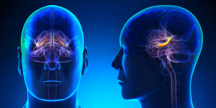

An analysis of more than 2,000 cognitively healthy older adults found that those with depression tended to have a smaller volume in one part of the hippocampus, a brain region central to memory and one of the first affected by Alzheimer’s disease. The association did not appear to be explained by amyloid and tau, two proteins linked to Alzheimer’s disease. The paper was published in Translational Psychiatry.
Alzheimer’s disease is the most common form of dementia in older adults. It gradually disrupts memory, thinking, behavior, and everyday functioning. The disease is accompanied by characteristic changes in the brain, including abnormal clumps of amyloid protein, called plaques, and twisted strands of tau protein, called tangles, along with the loss of nerve cells.
Research suggests that depression may also play a role in the risk of Alzheimer’s disease. People who experience depression during their lifetime appear to have a higher risk of developing Alzheimer’s disease later on. One possible connection between the two conditions involves the hippocampus, which is central to memory and is among the first areas affected by Alzheimer’s disease.
Depression has likewise been associated with changes in the structure of the hippocampus, although previous studies have produced inconsistent findings. Part of the difficulty may be that the hippocampus is not a single uniform structure but consists of several smaller regions (called hippocampal subfields) that can be affected differently by depression and Alzheimer’s disease. Antidepressant medications may also play a role, as some evidence suggests that they could protect against hippocampal shrinkage, while other findings raise questions about their effects in older adults.
Study author Danielle Luu of the Stevens Neuroimaging and Informatics Institute at the University of Southern California and colleagues set out to examine how depression and antidepressant use relate to the volume of hippocampal subfields in older adults without cognitive problems. They also wanted to know whether Alzheimer’s-related factors, including amyloid and tau buildup and the APOE4 gene variant, which raises Alzheimer’s risk, play a role in these relationships.
The researchers considered two possibilities for antidepressants. The medications might be linked to larger subfield volumes by relieving depressive symptoms. Alternatively, they might be linked to smaller volumes, either because of unknown harmful effects on the brain or because doctors are more likely to prescribe them to people with more severe depression.
The researchers analyzed data from the Health and Aging Brain Study-Health Disparities, which recruited Hispanic, Black, and white older adults through the University of North Texas in Fort Worth. The analysis included 2,009 cognitively unimpaired adults between 50 and 90 years of age, with an average age of about 65.
Of these participants, 630 were classified as having depression, either because they scored 10 or higher on the Geriatric Depression Scale or because they reported having been diagnosed with depression by a health care provider. The remaining 1,379 participants were classified as not depressed.
Participants underwent high-resolution MRI scans of the hippocampus and reported whether they used antidepressant medications. The researchers used the scans to calculate volumes of three hippocampal subfields: the CA1, the subiculum, and a composite region that included the CA2, CA3, and the dentate gyrus (CA23DG). Subsets of participants also underwent PET scans measuring amyloid (684 people) and tau (386 people) in the brain.
Participants with depression tended to have a smaller CA23DG, but depression was not associated with the volume of the CA1 or the subiculum. The association was small and held after the researchers accounted for body mass index and physical activity, as well as tau levels and APOE4 status. When amyloid was added to the analysis, the association was no longer statistically significant, but its size was similar, which the authors attribute to the smaller number of participants with amyloid scans.
The severity of current depressive symptoms and the age at which depression began were not associated with the volume of any subfield. In exploratory analyses, the link between depression and smaller subfields appeared stronger in men than in women, but the authors interpret this cautiously because the formal test of a sex difference was not statistically significant.
Among participants with depression, those who used antidepressant medications tended to have smaller CA1 and CA23DG volumes than those who did not. These associations weakened to borderline levels after the researchers accounted for body mass index and physical activity. When antidepressant use and depression were included in the same analysis, the link between depression itself and a smaller CA23DG faded, while antidepressant use remained associated with a smaller CA23DG.
The authors suggest that the medication findings may reflect what is known as indication bias, meaning that people with more severe depression may be more likely to be prescribed medication in the first place. Antidepressant users reported fewer current depressive symptoms than non-users, suggesting the medications were relieving symptoms, but the researchers did not have information on how severe participants’ depression was before treatment or how long they had taken medication.
“It is important to study AD [Alzheimer’s disease] in the context of mental health, since rates of depression rise with age. Moreover, it is crucial to investigate how depression and antidepressant medication use affect the hippocampus, a region of the brain that is heavily impacted by AD. Understanding the relationship between depression and the development of AD is important for exploring preventative measures against brain atrophy and dementia,” study authors concluded.
The study contributes to the scientific understanding of the neural underpinnings of depression in older adults. However, it should be noted that the design of this study does not allow any causal inferences to be derived from the results. Depression diagnoses and medication use were self-reported rather than drawn from medical records, and the analyses involving amyloid and tau were based on smaller subsets of participants.
The researchers did not compare different types or doses of antidepressants. They note that the possibility that the medications themselves affect hippocampal structure should be tested in randomized controlled trials.
The paper, “Depression and hippocampal subfield volume in older adults,” was authored by Danielle Luu, Alicia M. Twisselmann, Victoria R. Tennant, Brandon J. Hall, Arjun Mahajan, Audrey Kim, Jamie Terner, Marylan L. Davison, Koral Wheeler, Christopher R. K. Ching, James Hall, Matthew T. Borzage, Leigh Johnson, Arthur W. Toga, Sid E. O’Bryant, Kristine Yaffe, and Meredith N. Braskie for the HABS-HD Study Team.